. 2019;8(1):516-530.
doi: 10.1080/22221751.2019.1597644.

Towards a solution to MERS: protective human monoclonal antibodies targeting different domains and functions of the MERS-coronavirus spike glycoprotein

Affiliations

Towards a solution to MERS: protective human monoclonal antibodies targeting different domains and functions of the MERS-coronavirus spike glycoprotein

Ivy Widjaja et al. Emerg Microbes Infect. 2019.

Abstract

The Middle-East respiratory syndrome coronavirus (MERS-CoV) is a zoonotic virus that causes severe and often fatal respiratory disease in humans. Efforts to develop antibody-based therapies have focused on neutralizing antibodies that target the receptor binding domain of the viral spike protein thereby blocking receptor binding. Here, we developed a set of human monoclonal antibodies that target functionally distinct domains of the MERS-CoV spike protein. These antibodies belong to six distinct epitope groups and interfere with the three critical entry functions of the MERS-CoV spike protein: sialic acid binding, receptor binding and membrane fusion. Passive immunization with potently as well as with poorly neutralizing antibodies protected mice from lethal MERS-CoV challenge. Collectively, these antibodies offer new ways to gain humoral protection in humans against the emerging MERS-CoV by targeting different spike protein epitopes and functions.

Keywords: Coronavirus; MERS; antibodies; spike protein.


Figures
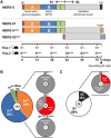
Figure 1.
Figure 1.
Generation and characterization of monoclonal antibodies targeting the MERS-CoV spike protein. (A) The MERS-CoV spike (S) protein and recombinant soluble MERS-CoV S antigens used for immunization of H2L2 transgenic mice to generate human monoclonal antibodies (mAbs). Upper panel: Schematic representation of the MERS-CoV S protein, indicated are S subunits (S1 and S2), S1 domains (A through D), and known biological functions. Middle panel: Schematic representation of recombinant soluble MERS-CoV S antigens, including the MERS-CoV S S1 subunit (MERS-S1), the ectodomain of its S2 subunit (MERS-S2ecto) or the entire MERS-S ectodomain (MERS-Secto), the latter containing a mutation at the furin cleavage site at the S1/S2 junction and a C-terminally fused T4 foldon trimerization tag to increase trimer stability (T4). Positions of signal peptides (SP) and StrepTag affinity tags (ST) are indicated. Lower panel: Immunization schedule H2L2 mice. To generate monoclonal antibodies (mAbs) targeting the MERS-CoV S protein, groups of H2L2 mice (six mice/group) were immunized with either MERS-S1 (6×), or sequentially immunized with MERS-Secto (3×), MERS-S2ecto (2×) and MERS-Secto (1x). Booster immunizations were done with two-week intervals and B-cells were harvested from spleen and lymph nodes four days after the last immunization. (B) Identified MERS-S1-reactive mAbs of hybridomas derived from B-cells of S1-immunized H2L2 mice were characterized for epitope location and virus neutralization using MERS-S pseudotyped VSV. Pie charts show mAb frequencies relative to the total (indicated in the centre circle). Domain-level epitope mapping was performed for MERS-S1-reactive mAbs and relative frequencies of mAbs binding to given S1 domains (S1A, S1B or S1CD) are indicated. The percentage of mAbs that was reactive to S1 but not to the S1A, S1B or S1CD domains (S1other) is also shown. Virus neutralization by S1-reactive mAbs was analysed using the luciferase-encoding MERS-CoV S pseudotyped VSV particles. (C) Identified MERS-Secto-reactive mAbs of hybridomas generated from Secto/S2ecto immunized H2L2 mice were characterized for epitope location and virus neutralization as in (B).
Figure 2.
Figure 2.
Human anti-MERS-S mAbs targeting six epitope groups distributed over multiple domains of the MERS-CoV spike protein. (A) ELISA reactivity of the human anti-MERS-S mAbs to the indicated MERS-CoV spike glycoprotein domains. (B) Binding competition of anti-MERS-S mAbs analysed by bio-layer interferometry (BLI). Immobilized MERS-Secto antigen was saturated in binding with a given anti-MERS-S mAb (step 1) and then exposed to binding by a second mAb (step 2). Additional binding of the second antibody indicates the presence of an unoccupied epitope, whereas lack of binding indicates epitope blocking by the first antibody. As a control, the first mAb was also included in the second step to check for self-competition. (C) Schematic distribution of epitope groups of anti-MERS-S mAbs over the different MERS-S domains.
Figure 3.
Figure 3.
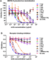
Virus neutralization and receptor binding inhibition by anti-MERS-S mAbs. (A) Analysis of MERS-CoV neutralizing activity by anti-MERS-S mAbs using MERS-S pseudotyped, luciferase-encoding VSV. A previously described RBD-specific, MERS-CoV-neutralizing human monoclonal antibody (anti-MERS-CTRL) and irrelevant isotype monoclonal antibody (Iso-CTRL) were included as positive and negative control, respectively. Luciferase-expressing VSV particles pseudotyped with the MERS-CoV S protein or authentic MERS-CoV were incubated with antibodies at the indicated concentrations and the mix was used to transduce Huh-7 cells. At 24 h postinfection luciferase expression was measured and neutralization (%) was calculated as the ratio of luciferase signal relative to relative to non-antibody-treated controls. Data represent the mean (± standard deviation, SD) of three independent experiments. (B) Receptor binding inhibition by anti-MERS-S mAbs, determined by an ELISA-based assay. Recombinant soluble MERS-Secto was preincubated with serially diluted anti-MERS-S mAbs and added to ELISA plates coated with soluble the MERS-CoV S ectodomain. Binding of MERS-Secto to DPP4 was measured using HRP-conjugated antibody recognizing the Streptag affinity tag on DPP4. Data represent the mean (± standard deviation, SD) of three independent experiments.
Figure 4.
Figure 4.
Anti-MERS-S mAbs targeting MERS-S1A and -S2 domains block domain-specific functions. (A) The anti-MERS-S1A mAb 1.10f3 interferes with MERS-S1A-mediated sialic acid binding, determined by a hemagglutination inhibition assay [16]. The sialic-acid binding domain S1A of MERS-S was fused to lumazine synthase (LS) protein that can self-assemble to form 60-meric nanoparticle (S1A-LS), which enables multivalent, high affinity binding of the MERS-S1A domain to sialic acid ligands such as on erythocytes. Human red blood cells were mixed with S1A-LS in the absence or presence of 2-fold dilutions of the MERS-S1A-specific mAb 1.10f3. Isotype control antibody was included as a negative control. Hemagglutination was scored after 2 h of incubation at 4°C. The hemagglutination inhibition assay was performed three times, a representative experiment is shown. (B) Neutralization of MERS-S pseudotyped VSV by anti-MERS-S mAb 1.10f3 on Vero and Calu-3 cells. Data represent the mean (± standard deviation, SD) of three independent experiments. (C) The anti-MERS-S2 mAbs 1.6c7 and 3.5g6 block MERS-S-mediated cell–cell fusion. Huh-7 cells were transfected with plasmid expressing MERS-CoV S, C-terminally fused to GFP. Two days after transfection, cells were treated with trypsin to activate membrane the fusion function of the MERS-CoV S protein, and incubated in the presence or absence of anti-MERS-S2 mAbs 1.6c7 and 3.5g6, or the anti-MERS-S1B mAb 7.7g6 and anti-MERS-S1A 1.10f3, all at 10 μg/ml. Formation of MERS-S mediated cell–cell fusion was visualized by fluorescence microscopy. Merged images of MERS-S-GFP expressing cells (green) and DAPI-stained cell nuclei (blue) are shown. Experiment was repeated two times and representative images are shown.
Figure 5.
Figure 5.
Human anti-MERS-S mAbs protect mice against lethal MERS-CoV challenge. (A–B) Fifty microgram of antibody (equivalent to 1.8 mg mAb/kg body weight) was infused intraperitoneally in K18-hDPP4-transgenic mice 6 h before challenge with 5 × 103 pfu/mice of MERS-CoV. Five mice per group were used in the experiment. Survival rates (A) and weight loss (B) (expressed as a percentage of the initial weight) was monitored daily until 12 days post-inoculation.

References

    1. http://www.emro.who.int/health-topics/mers-cov/mers-outbreaks.html.
    1. Omrani A. S., Al-Tawfiq J. A., and Memish Z. A.. 2015. Middle east respiratory syndrome coronavirus (MERS-CoV): animal to human interaction. Pathog. Glob. Health. 109:354–362. doi:10.1080/20477724.2015.1122852. - DOI - PMC - PubMed
    1. Azhar E. I., El-Kafrawy S. A., Farraj S. A., Hassan A. M., Al-Saeed M. S., Hashem A. M., and Madani T. A.. 2014. Evidence for camel-to-human transmission of MERS coronavirus. N. Engl. J. Med. 370:2499–2505. doi:10.1056/NEJMoa1401505. - DOI - PubMed
    1. Haagmans B. L., Al Dhahiry S. H., Reusken C. B., Raj V. S., Galiano M., Myers R., Godeke G. J., Jonges M., Farag E., Diab A., Ghobashy H., Alhajri F., Al-Thani M., Al-Marri S. A., Al Romaihi H. E., Al Khal A., Bermingham A., Osterhaus A. D., AlHajri M. M., and Koopmans M. P.. 2014. Middle East respiratory syndrome coronavirus in dromedary camels: an outbreak investigation. Lancet Infect. Dis. 14:140–145. doi:10.1016/S1473-3099(13)70690-X. - DOI - PMC - PubMed
    1. Memish Z. A., Cotten M., Meyer B., Watson S. J., Alsahafi A. J., Al Rabeeah A. A., Corman V. M., Sieberg A., Makhdoom H. Q., Assiri A., Al Masri M., Aldabbagh S., Bosch B. J., Beer M., Muller M. A., Kellam P., and Drosten C.. 2014. Human infection with MERS coronavirus after exposure to infected camels, Saudi Arabia, 2013. Emerg Infect Dis 20:1012–1015. doi:10.3201/eid2006.140402. - DOI - PMC - PubMed

MeSH terms