. 2019 Apr 2;14(4):e0214476.
doi: 10.1371/journal.pone.0214476. eCollection 2019.

Sodium thiocyanate treatment attenuates atherosclerotic plaque formation and improves endothelial regeneration in mice

Affiliations

Sodium thiocyanate treatment attenuates atherosclerotic plaque formation and improves endothelial regeneration in mice

Andreas Zietzer et al. PLoS One. .

Abstract

Introduction: Atherosclerotic plaque formation is an inflammatory process that involves the recruitment of neutrophil granulocytes and the generation of reactive oxygen species (ROS). ROS formation by myeloperoxidase, a key enzyme in H2O2 degradation, can be modulated by addition of sodium thiocyanate (NaSCN). However, the therapeutic use of NaSCN to counteract atherogenesis has been controversial, because MPO oxidizes NaSCN to hypothiocyanous acid, which is a reactive oxygen species itself. Therefore, this study aimed to investigate the effect of NaSCN treatment on atherogenesis in vivo.

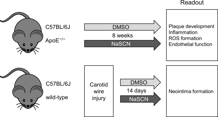
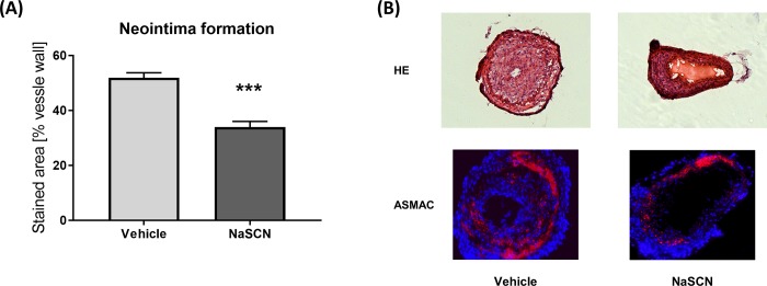
Methods: Apolipoprotein E knockout (ApoE-/-) mice on western-diet were treated with NaSCN for 8 weeks. Blood levels of total cholesterol, IL-10, and IL-6 were measured. Aortic roots from these mice were analyzed histologically to quantify plaque formation, monocyte, and neutrophil granulocyte infiltration. Oxidative damage was evaluated via an L-012 chemiluminescence assay and staining for chlorotyrosine in the aortic walls. Endothelial function was assessed by use of endothelium-dependent vasodilation in isolated aortic rings. Neointima formation was evaluated in wild-type mice following wire injury of the carotid artery.

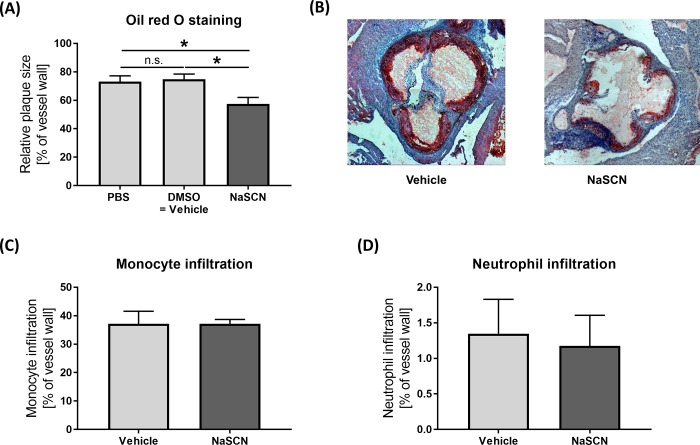
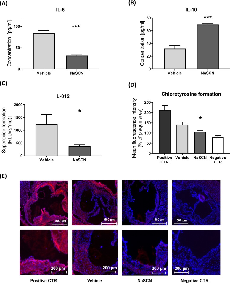
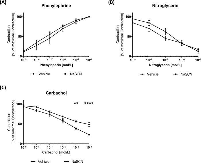
Results: NaSCN treatment of ApoE-/- mice lead to a reduction of atherosclerotic plaque size in the aortic roots but had no effect on monocyte or granulocyte infiltration. Serum levels of the pro-inflammatory cytokine IL-6 decreased whereas anti-inflammatory IL-10 increased upon NaSCN treatment. In our experiments, we found oxidative damage to be reduced and the endothelial function to be improved in the NaSCN-treated group. Additionally, NaSCN inhibited neointima formation.

Conclusion: NaSCN has beneficial effects on various stages of atherosclerotic plaque development in mice.


Conflict of interest statement

The authors have declared that no competing interests exist.

Figures

Fig 1
Fig 1. Schematic illustration of the experimental design.
For the evaluation of plaque development, inflammation, ROS formation, and endothelial function ApoE-/- mice were used after 8 weeks of intraperitoneal application of NaSCN (200 μg every other day) or vehicle (DMSO). For the assessment of neointima formation wild-type mice were subjected to carotid artery wire injury, treated with NaSCN (200 μg every other day) or vehicle for 14 days, and then analyzed.
Fig 2
Fig 2. Assessment of atherosclerotic plaque formation, monocyte, and neutrophil granulocyte infiltration in ApoE-/- mice upon NaSCN treatment or DMSO as vehicle.
(A) Quantitative analysis of plaque size as a percentage of the total aortic root vessel wall by oil red staining, n = 5–6. (B) Representative histological images of the aortic root (oil red + hematoxylin staining). (C + D) Histological assessment of monocyte and neutrophil granulocyte infiltration as a percentage of the total vessel wall by immunohistological staining, n = 5. Data are presented as the mean ± SEM., *p ≤ 0.05, ***p ≤ 0.005 vs. vehicle.
Fig 3
Fig 3. Assessment of IL-6 and IL-10 plasma levels and ROS / chlorotyrosine formation in ApoE-/- mice upon NaSCN treatment or DMSO as vehicle.
(A + B) Plasma IL-6 and IL-10 levels upon NaSCN treatment measured by ELISA, n = 4. (C) Measurement of ROS formation in aortic segments by L-012 chemiluminescence, n = 4. (D) Quantification of HOCl-dependent tissue damage via immunohistological staining of 3-cholortyrosine in the atherosclerotic plaque area, n = 4. (E) Representative images of Chlorotyrosine staining, positive control after incubation with 0.005% HOCl for 1 hour, negative control only with secondary anti-body (Red chlorotyrosine, Blue DAPI). Data are presented as the mean ± SEM., n = 4, *p ≤ 0.05, ***p ≤ 0.005 vs. vehicle.
Fig 4
Fig 4. Measurement of endothelial function in isolated aortic segments of ApoE−/− mice upon NaSCN treatment in organ chamber experiments.
(A) Definition of the maximal endothelial contraction by incubation with increasing phenylephrine concentrations. (B) Assessment of endothelium-independent vasodilation as a percentage of the maximal contraction with increasing concentrations of nitroglycerin. (C) Assessment of endothelium-dependent vasodilation as a percentage of the maximal contraction with increasing concentrations of carbachol. Data are presented as the mean ± SEM, n = 4–5, **p ≤ 0.01, ***p ≤ 0.005 vs. vehicle.
Fig 5
Fig 5. Evaluation of neointima formation after carotid-artery wire injury in wild-type mice upon NaSCN treatment.
(A) Quantitative analysis of neointima formation by use of hematoxylin/eosin staining as a percentage of the vessel wall. (B) Representative histological images of the injured carotid artery 14 days post injury. Upper panel with hematoxylin/eosin staining. Lower panel with anti-α-smooth-muscle Actin staining (red) and DAPI (blue). Data are presented as the mean ± SEM, n = 5, ***p ≤ 0.005 vs. vehicle.

References

    1. The World Health Organization. Global Health Estimates 2016: Deaths by Cause, Age, Sex, by Country and by Region, 2000–2016 Geneva, World Health Organization; 2018. Available from: https://www.who.int/healthinfo/global_burden_disease/estimates/en/index1...
    1. Pasceri V, Cheng JS, Willerson JT, Yeh ET, Chang J. Modulation of C-reactive protein-mediated monocyte chemoattractant protein-1 induction in human endothelial cells by anti-atherosclerosis drugs. Circulation. 2001; 103: 2531–2534. - PubMed
    1. Libby P, Ridker PM, Hansson GK. Progress and challenges in translating the biology of atherosclerosis. Nature. 2011; 473: 317–325. 10.1038/nature10146 - DOI - PubMed
    1. Lau D, Baldus S. Myeloperoxidase and its contributory role in inflammatory vascular disease. Pharmacol Ther. 2006; 111: 16–26. 10.1016/j.pharmthera.2005.06.023 - DOI - PubMed
    1. Ronald JA, Chen JW, Chen Y, Hamilton AM, Rodriguez E, Reynolds F, et al. Enzyme-sensitive magnetic resonance imaging targeting myeloperoxidase identifies active inflammation in experimental rabbit atherosclerotic plaques. Circulation. 2009; 120: 592–599. 10.1161/CIRCULATIONAHA.108.813998 - DOI - PMC - PubMed

Publication types

MeSH terms