. 2019 Apr 2;19(1):85.
doi: 10.1186/s12886-019-1091-4.

Individualized penetrating keratoplasty using edge-trimmed glycerol-preserved donor corneas for perforated corneal ulcers

Affiliations

Individualized penetrating keratoplasty using edge-trimmed glycerol-preserved donor corneas for perforated corneal ulcers

Guozhen Niu et al. BMC Ophthalmol. .

Abstract

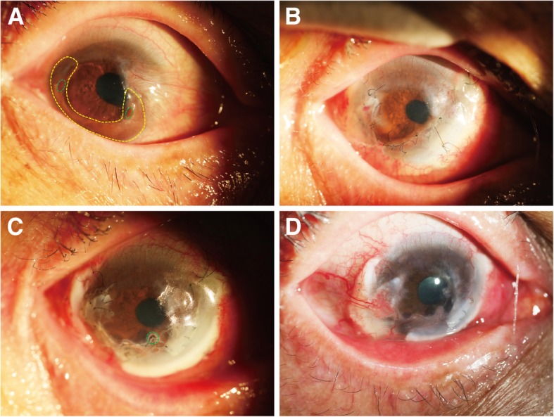
Purpose: To report a surgical technique and the surgical outcomes of individualized penetrating keratoplasty (PK) using edge-trimmed glycerol-preserved donor corneas for perforated corneal ulcers.

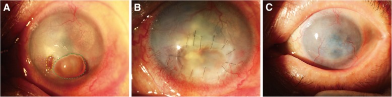
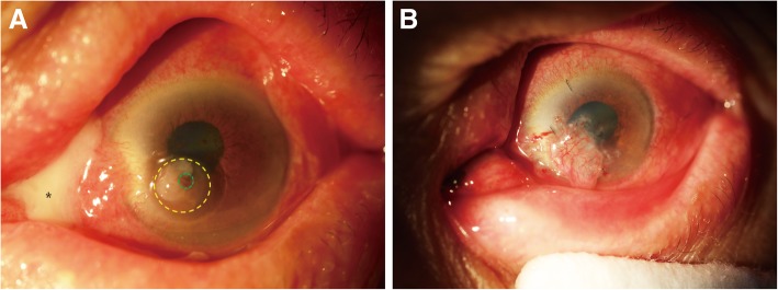
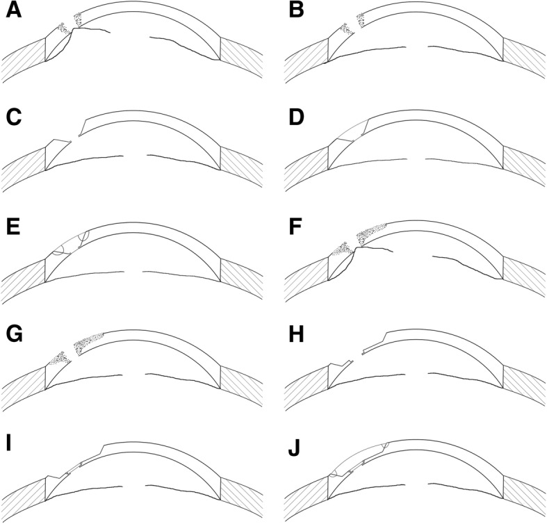
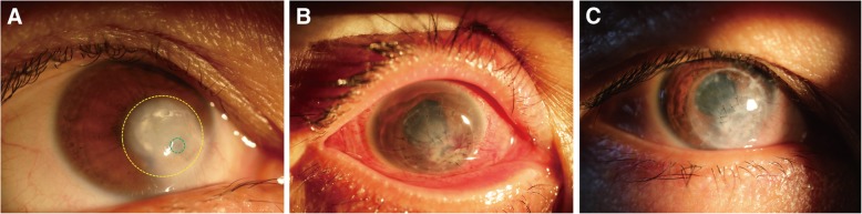
Methods: Fourteen perforated eyes from 14 patients who underwent individualized PK using edge-trimmed glycerol-preserved donor corneas, were included in the retrospective study. The perforations were mainly 1-2 mm in size except for one that was 2.5 × 4 mm. Three patients were treated with PK; one patient was treated with PK and a conjunctival flap; ten patients who had large ulcer areas were treated with PK combined with lamellar keratoplasty (LK). Donor corneas were preserved in sterile pure glycerol at - 80 °C. Corneal grafts were specially edge-trimmed to match the perforation, and then sutured onto the recipient bed avoiding the visual axis.

Results: All 14 patients recovered anatomical integrity without reinfections of the treated eyes. All patients had improved graft transparency and uncorrected visual acuity after surgery. Among them, four patients suffered from short-term postoperative complications and recovered quickly; four patients suffered from long-term postoperative complications, of them, one was performed further treatment.

Conclusion: After individualized PK using glycerol-preserved donor corneas, all perforated corneal ulcers were stably controlled by the end of the follow-up period. This modified surgical technique can be a potential treatment choice for patients with perforated corneal ulcers.

Keywords: Cornea; Corneal perforation; Individualized penetrating keratoplasty; Preserved.


Conflict of interest statement

Ethics approval and consent to participate

This study was approved by the Ethics Committee of Tongji Hospital, Tongji University School of Medicine, and adhered to the tenets of the Declaration of Helsinki. Due to the retrospective nature of the study, informed consent was waived.

Consent for publication

Written informed consent was obtained from the patients for publication of this article and any accompanying images. Copies of the written consents are available for review by the Editor of this journal.

Competing interests

The authors declare that they have no competing interests.

Publisher’s Note

Springer Nature remains neutral with regard to jurisdictional claims in published maps and institutional affiliations.

Figures

Fig. 1
Fig. 1
Patient 5 (necrotizing stromal keratitis (HSK), visual acuity: HM, left eye). a Green dotted circle indicates the perforation (4 × 2.5 mm in diameter) with iris prolapse; yellow dotted circle indicates a small corneal ulcer beside the perforation. b An edge-trimmed PK graft covered both perforated and ulcerated regions was sutured onto the recipient bed. c 16 months after surgery, the graft was stable, with improved transparency
Fig. 2
Fig. 2
Patient 1 (upper lacrimal duct inflammation, ophthalmodynia and massive secretions, left eye). a Green dotted circle indicates the perforation (1 × 1 mm in diameter) with iris prolapse; yellow dotted circle indicates a corneal ulcer; asterisk indicates a large amount of white purulent secretion emitted from the upper lacrimal punctum. b After edge-trimmed PK surgery, a conjunctival flap was used to cover the corneal graft to avoid corneal reinfection
Fig. 3
Fig. 3
Schematic diagrams for surgical steps. a Perforation with iris prolapse and shallow anterior chamber. b Iris and anterior chamber restored after Healon injection. c The recipient bed was produced with sloping surface by excising the necrotic tissue and trimming the perforation edge. d Trimmed corneal graft with wedged edge was inserted into the recipient bed. e Watertight suture. f Perforation with iris prolapse, shallow anterior chamber and large corneal ulcer. g Iris and anterior chamber restored after Healon injection. h The recipient bed was produced by excising the necrotic tissue and trimming the perforation edge. i A thin PK graft (approximately 100 μm) was transplanted to the perforated area with watertight suture. j A thicker and larger-edged trimmed LK graft was sutured onto the region of the corneal ulcer
Fig. 4
Fig. 4
Patient 3 (noninfectious, visual acuity: LP, right eye). a Green dotted circle indicates the perforation (1.5 × 1.5 mm in diameter) with a small area of iris prolapse; yellow dotted circle indicates a large circular area of corneal ulcer and leucoma. b A small, thin PK graft was sutured to close the perforation, then a LK graft—larger than the ulcerated region—was sutured across the optical axis for a better visual acuity after surgery. c 9 months after surgery, the graft showed improved transparency
Fig. 5
Fig. 5
Patient 7 (Mooren’s ulcer, visual acuity: CF, right eye). a Green dotted circle indicates two perforations (1 × 1 mm in diameter) with prolapsed iris; yellow dotted circle indicates a large crescent ulcer at the peripheral region; a pterygium was at the nasal side. b The pterygium was excised, and individualized PK was performed with two thin, penetrating corneal grafts to cover the perforations. A crescent LK graft covered the PK grafts and the ulcerated areas around the perforations, including the region where pterygium was excised. c Green dotted circle indicates a micro-perforation (1.5 months after the surgery), which was cured by the transplantation of a conjunctival flap. d After a follow-up period of 13 months, the graft showed improved transparency

Similar articles

Cited by

References

    1. Boruchoff SA, Donshik PC. Medical and surgical management of corneal thinnings and perforations. Int Ophthalmol Clin. 1975;15(4):111–123. doi: 10.1097/00004397-197501540-00010. - DOI - PubMed
    1. Portnoy SL, Insler MS, Kaufman HE. Surgical management of corneal ulceration and perforation. Surv Ophthalmol. 1989;34(1):47–58. doi: 10.1016/0039-6257(89)90129-X. - DOI - PubMed
    1. Jhanji V, Young AL, Mehta JS, Sharma N, Agarwal T, Vajpayee RB. Management of corneal perforation. Surv Ophthalmol. 2011;56(6):522–538. doi: 10.1016/j.survophthal.2011.06.003. - DOI - PubMed
    1. Krysik K, Dobrowolski D, Lyssek-Boron A, Jankowska-Szmul J, Wylegala EA. Differences in surgical Management of Corneal Perforations, measured over six years. J Ophthalmol. 2017;2017:1582532. - PMC - PubMed
    1. Poon AC, Geerling G, Dart JK, Fraenkel GE, Daniels JT. Autologous serum eyedrops for dry eyes and epithelial defects: clinical and in vitro toxicity studies. Br J Ophthalmol. 2001;85(10):1188–1197. doi: 10.1136/bjo.85.10.1188. - DOI - PMC - PubMed

LinkOut - more resources