

Exosomal let-7d-3p and miR-30d-5p as diagnostic biomarkers for non-invasive screening of cervical cancer and its precursors
- PMID: 30940131
- PMCID: PMC6446401
- DOI: 10.1186/s12943-019-0999-x
Exosomal let-7d-3p and miR-30d-5p as diagnostic biomarkers for non-invasive screening of cervical cancer and its precursors
Abstract
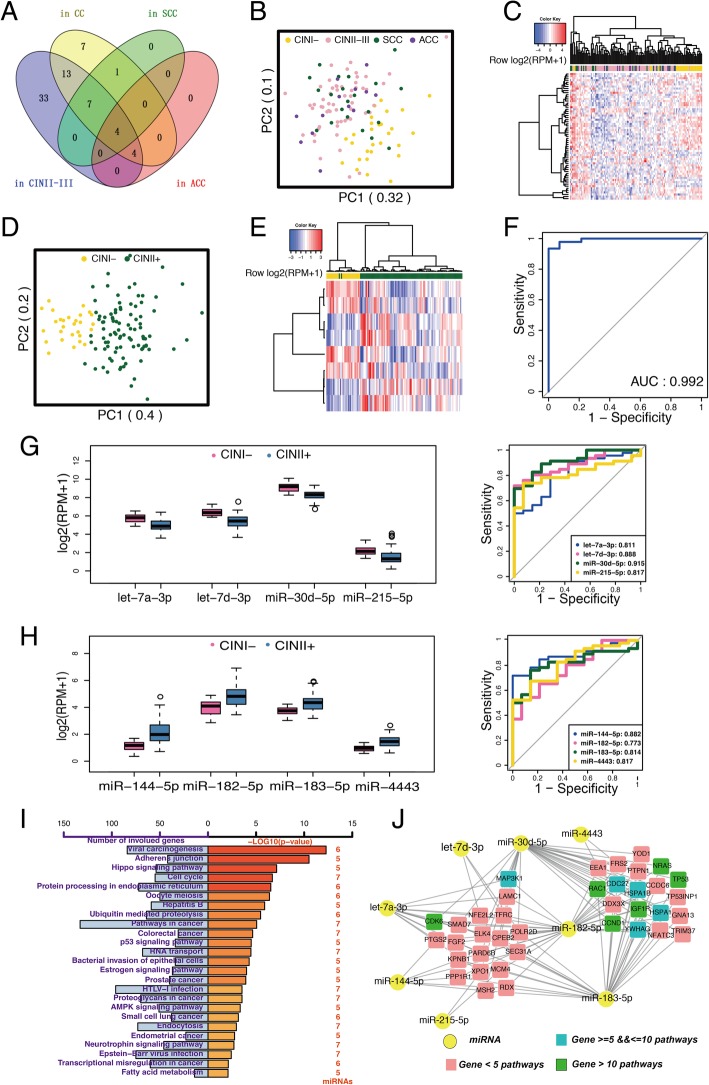
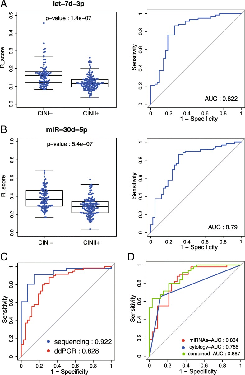
Cervical cancer screening through detection and treatment of high-grade cervical intraepithelial neoplasia (CIN) is most successful in cancer prevention. However, the accuracy of the current cervical cancer screening tests is still low. The aim of this study was to develop a more accurate method based on circulating exosomal miRNAs. The miRNA sequencing was performed to identify candidate exosomal miRNAs as diagnostic biomarkers in 121 plasma samples from healthy volunteers, cervical carcinoma patients, and CIN patients. A panel with eight differentially expressed exosomal miRNAs was identified to distinguish patients in the CIN II+ group (including advanced CIN II patients) from those in the CIN I- group (including CIN I patients and healthy volunteers). Let-7d-3p and miR-30d-5p showed significant difference between cervical tumors and adjacent normal tissues (P < 0.005), exhibited a consistent trend in plasma samples, and were further validated in 203 independent plasma samples. Integrating these two miRNAs yielded an AUC value of 0.828 to distinguish patients in CIN II+ group from those in CIN I- group. Further integrating them into a cytological test-based model resulted in a higher AUC of 0.887, while the AUC value based on the cytological test alone was 0.766. In summary, plasma exosomal miR-30d-5p and let-7d-3p are valuable diagnostic biomarkers for non-invasive screening of cervical cancer and its precursors. Further validation using large sample sizes is required for clinical diagnosis.
Keywords: Cervical cancer; Diagnosis; Early detection; Exosome; Liquid biopsy; Next-generation sequencing; miRNA.
Conflict of interest statement
Ethics approval and consent to participate
This study was reviewed and approved by the Ethnics Committees of Women’s Hospital of Zhejiang University School of Medicine and Tongde Hospital of Zhejiang Province (Hangzhou, China). The study was conducted in accordance with the International Ethical Guidelines for Biomedical Research Involving Human Subjects.
Consent for publication
All authors have agreed to publish this manuscript.
Competing interests
The authors declare that they have no competing interests.
Publisher’s Note
Springer Nature remains neutral with regard to jurisdictional claims in published maps and institutional affiliations.
Figures
References
-
- Chen WQ, Zheng RS, Baade PD, Zhang SW, Zeng HM, Bray F, Jemal A, Yu XQ, He J: Cancer Statistics in China, 2015. Ca-a Cancer Journal for Clinicians 2016;66:115–32. - PubMed
-
- Smith RA, Manassaram-Baptiste D, Brooks D, Doroshenk M, Fedewa S, Saslow D, Brawley OW, Wender R: Cancer Screening in the United States, 2015: A Review of Current American Cancer Society Guidelines and Current Issues in Cancer Screening. Ca-a Cancer Journal for Clinicians 2015;65:30–54. - PubMed
-
- Denzer K, Kleijmeer MJ, Heijnen HFG, Stoorvogel W, Geuze HJ: Exosome: from internal vesicle of the multivesicular body to intercellular signaling device. Journal of Cell Science 2000;113:3365–74. - PubMed
-
- Taylor DD, Gercel-Taylor C: MicroRNA signatures of tumor-derived exosomes as diagnostic biomarkers of ovarian cancer. Gynecologic Oncology 2008;110:13–21. - PubMed
