. 2019 Apr 9;3(7):1039-1046.
doi: 10.1182/bloodadvances.2018030635.

Germline deletion of ETV6 in familial acute lymphoblastic leukemia

Affiliations

Germline deletion of ETV6 in familial acute lymphoblastic leukemia

Evadnie Rampersaud et al. Blood Adv. .

Abstract

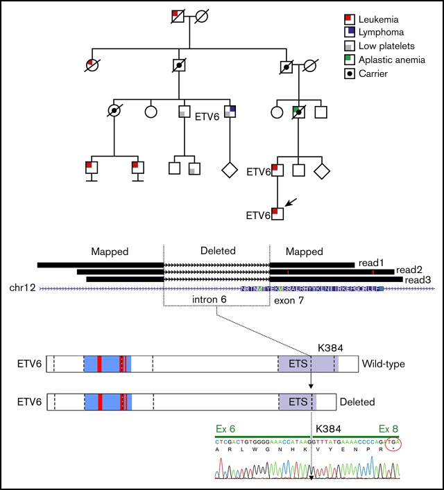
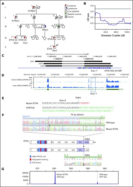
Recent studies have identified germline mutations in TP53, PAX5, ETV6, and IKZF1 in kindreds with familial acute lymphoblastic leukemia (ALL), but the genetic basis of ALL in many kindreds is unknown despite mutational analysis of the exome. Here, we report a germline deletion of ETV6 identified by linkage and structural variant analysis of whole-genome sequencing data segregating in a kindred with thrombocytopenia, B-progenitor acute lymphoblastic leukemia, and diffuse large B-cell lymphoma. The 75-nt deletion removed the ETV6 exon 7 splice acceptor, resulting in exon skipping and protein truncation. The ETV6 deletion was also identified by optimal structural variant analysis of exome sequencing data. These findings identify a new mechanism of germline predisposition in ALL and implicate ETV6 germline variation in predisposition to lymphoma. Importantly, these data highlight the importance of germline structural variant analysis in the search for germline variants predisposing to familial leukemia.


Conflict of interest statement

Conflict-of-interest disclosure: C.G.M. has received consulting fees and travel funding from Amgen and Pfizer and research funding from AbbVie, Loxo Oncology, and Pfizer. The content of these activities and research is unrelated to the content of this manuscript. The remaining authors declare no competing financial interests.

Figures

None
Graphical abstract
Figure 1.
Figure 1.
Linkage mapping to chromosome 12 in a kindred with ALL and DLBCL. (A) A 5-generation kindred with 10 individuals with leukemia, DLBCL, aplastic anemia, and/or thrombocytopenia. Black crosses indicate samples subjected to WGS, and red crosses indicate samples subjected to WES. Squares and circles represent male and females, respectively. All family members with ALL or DLBCL sequenced harbored the ETV6 deletion. Individuals III.3 and III.4 (DLBCL) both had thrombocytopenia and the deletion; IV.3 and III.2 had normal platelet counts and no ETV6 deletion. (B) Multipoint linkage results highlighting region with a logarithm of the odds (LOD) score of 1.8. (C) Representative reads showing the exon 7 splice site deletion identified by WGS SV analysis (NM_001987.4:G.11885871_11885946del;NM_001987.4:c.1153-55_1173del). (D) RNA-sequencing coverage analysis showing reduced sequence depth at exon 7. (E) Protein truncation resulting from exon skipping. (F) Schematic representation of the ETV6 protein, including the sterile α motif (SAM)/pointed domain of Tel/Yan protein, the polypeptide -binding domain, and the erythroblast transformation-specific domain (ETS). The top pair of electropherograms is from genomic PCR and Sanger sequencing and compares wild-type (WT) and deleted (75-bp) ETV6. Dotted line in the ETV6 cartoon represents exon junctions. The lower electropherogram is from RT-PCR and Sanger sequencing and shows the skipping of exon 7 and splicing of exon 6 to exon 8 in tumor RNA. (G) Results from fragment size analysis showing amplification of both WT and deleted ETV6 alleles.
Figure 2.
Figure 2.
Characterization of tumors in the ETV6-mutated kindred. (A) Immunohistochemistry of the DLBCL tumor is consistent with mature B-cell lineage, with expression of CD20 and lack of expression of the immature markers CD34 and TdT and the T-cell marker CD3, which highlights admixed small T cells (original magnification ×40; scale bars, 50 μm). (B) Mutational analysis of WGS data showing distinct ALL and DLBCL mutational spectra of each sample. (C) Gene set enrichment analysis (GSEA) from RNA sequencing of tumor sample showing enrichment of B lymphoid progenitor genes in the B-cell ALL sample (D) and genes expressed in lymphoma in the DLBCL sample. Collectively, the pathologic and genomic features support a true DLBCL in case III.4 rather than a lymphomatous presentation of ALL. Tdt, terminal deoxynucleotidyltransferase.
Figure 3.
Figure 3.
Workflow showing optimal practices for identification of germline variants predisposing to familial leukemia, incorporating analysis of soft-clipped reads. BWA, Burrows-Wheeler Aligner; LOD, logarithm of the odds.

References

    1. Iacobucci I, Mullighan CG. Genetic basis of acute lymphoblastic leukemia. J Clin Oncol. 2017;35(9):975-983. - PMC - PubMed
    1. Mullighan CG, Goorha S, Radtke I, et al. . Genome-wide analysis of genetic alterations in acute lymphoblastic leukaemia. Nature. 2007;446(7137):758-764. - PubMed
    1. Moriyama T, Relling MV, Yang JJ. Inherited genetic variation in childhood acute lymphoblastic leukemia. Blood. 2015;125(26):3988-3995. - PMC - PubMed
    1. Holmfeldt L, Wei L, Diaz-Flores E, et al. . The genomic landscape of hypodiploid acute lymphoblastic leukemia. Nat Genet. 2013;45(3):242-252. - PMC - PubMed
    1. Shah S, Schrader KA, Waanders E, et al. . A recurrent germline PAX5 mutation confers susceptibility to pre-B cell acute lymphoblastic leukemia. Nat Genet. 2013;45(10):1226-1231. - PMC - PubMed

Publication types

MeSH terms

Substances