. 2019 Jul 1;25(13):4079-4090.
doi: 10.1158/1078-0432.CCR-18-3223. Epub 2019 Apr 2.

Targeting Glutamine Metabolism and Redox State for Leukemia Therapy

Affiliations

Targeting Glutamine Metabolism and Redox State for Leukemia Therapy

Mark A Gregory et al. Clin Cancer Res. .

Abstract

Purpose: Acute myeloid leukemia (AML) is a hematologic malignancy characterized by the accumulation of immature myeloid precursor cells. AML is poorly responsive to conventional chemotherapy and a diagnosis of AML is usually fatal. More effective and less toxic forms of therapy are desperately needed. AML cells are known to be highly dependent on the amino acid glutamine for their survival. These studies were directed at determining the effects of glutaminase inhibition on metabolism in AML and identifying general weaknesses that can be exploited therapeutically.

Experimental design: AML cancer cell lines, primary AML cells, and mouse models of AML and acute lymphoblastic leukemia (ALL) were utilized.

Results: We show that blocking glutamine metabolism through the use of a glutaminase inhibitor (CB-839) significantly impairs antioxidant glutathione production in multiple types of AML, resulting in accretion of mitochondrial reactive oxygen species (mitoROS) and apoptotic cell death. Moreover, glutaminase inhibition makes AML cells susceptible to adjuvant drugs that further perturb mitochondrial redox state, such as arsenic trioxide (ATO) and homoharringtonine (HHT). Indeed, the combination of ATO or HHT with CB-839 exacerbates mitoROS and apoptosis, and leads to more complete cell death in AML cell lines, primary AML patient samples, and in vivo using mouse models of AML. In addition, these redox-targeted combination therapies are effective in eradicating ALL cells in vitro and in vivo.

Conclusions: Targeting glutamine metabolism in combination with drugs that perturb mitochondrial redox state represents an effective and potentially widely applicable therapeutic strategy for treating multiple types of leukemia.


Conflict of interest statement

Conflicts of Interest

The authors declare no conflicts of interest.

Figures
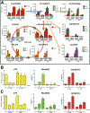
Figure 1.
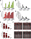
Figure 1.. The glutaminase inhibitor CB-839 impairs energy and redox metabolism in FLT3WT AML cells.
A) Glutamine flux analysis of OCI-AML3 and EOL-1 AML cells treated with vehicle (DMSO) or CB-839 (500 nM). Cells were pre-treated for 8 h followed by 13C,15N-glutamine labeling for 0, 3, 6, and 12 h, as indicated, followed by UHPLC/MS analysis. Levels of 13C and15N containing isotopologues (based on signal intensity) are shown. M+X+Y = molecular ion peak + no. of labeled 13C + no. of labeled 15N. B,C) EOL-1 cells were treated with CB-839 (0, 0.5, or 1 μM, as indicated) with simultaneous addition of cell-permeable α-ketoglutarate (4 mM α-KG; B) or cell-permeable glutathione (4 mM GSH; C) for 24 h and ATP levels and % of cells positive for MitoROS (detected using the dye MitoPY1) were measured. Apoptosis was measured after 48 h. For B and C, asterisks indicate statistical significance for comparisons of α-KG or GSH treated cells with control cells (0 α-KG or GSH) under the same CB-839 treatment conditions.
Figure 2.
Figure 2.. The glutaminase inhibitor CB-839 decreases glutathione levels, and induces mitoROS and apoptosis in AML cells.
A,B,C) The AML cell lines EOL-1, Molm13, MonoMac6, MV4–11, HL-60, Kasumi-1, OCI-AML3, and THP-1, and the lymphoma-derived cell line U937 and the CML cell lines K562 and KU812 were treated with CB-839 (0, 0.5, or 1 μM, as indicated) and glutathione (A) and mitoROS (B) were measured after 24 h using MitoPY1. Apoptosis was measured after 48 h (C). D) Model showing mechanism of redox-directed combination therapy for treating leukemia. Use of a glutaminase inhibitor (Drug 1, e.g. CB-839) blocks glutamine metabolism and production of antioxidant GSH and makes leukemia cells vulnerable to a pro-oxidant agent (Drug 2, e.g. ATO or HHT), leading to superinduction of mitoROS and subsequent leukemia cell death via apoptosis. For A, B, and C, asterisks indicate statistical significance for comparisons of CB-839 treated cells to untreated cells (0 CB).
Figure 3.
Figure 3.. CB-839 cooperates with the pro-oxidant drug ATO in inducing mitoROS, apoptosis, and AML cell elimination in vitro and in vivo.
EOL-1 (A,B,C) or Molm13 (D,E,F) were treated with CB-839 and ATO alone or together as indicated (increasing concentrations of ATO of 0, 0.2, 0.4, and 0.8 μM indicated by triangles) and mitoROS were measured using MitoPY1 (A,D). After 72 h, the number of viable cells (based on PI exclusion) was counted by flow cytometry (B,E) and apoptosis was measured after 48 h (C,F). G,H) MLL-ENL/FLT3-ITD+ luciferase expressing mouse leukemia cells were transplanted into Bl6 mice and after 3 days treatment was initiated with vehicle (n=5), CB-839 (n=5; 200 mg/kg twice daily p.o.), ATO (n=5; 8 mg/kg once daily i.p.), or CB-839 and ATO in combination (n=5) for up to 11 days (4 days on/2 days off/ 5 days on therapy). Leukemic burden was measured by bioluminescence using an in vivo imaging system (IVIS) on the indicated days (G). H) IVIS images from Day 11. For A, C, D, and F, asterisks indicate statistical significance for comparisons of CB-839 treated cells with control cells (0 CB) under the same ATO treatment conditions.
Figure 4.
Figure 4.. HHT induces mitoROS and cooperates with CB-839 in eliminating AML cells in vitro and in vivo.
A,B,C) Molm13 cells were treated with CB-839 and HHT alone or together as indicated for 20 h (triangles: 0, 5, 10, and 20 nM HHT) and mitoROS were measured using MitoPY1 (A). After 48h, the number of viable cells (based on PI exclusion) was counted by flow cytometry (B) and apoptosis was measured after 24 h (C). D) EOL-1 cells were treated with CB-839 and HHT alone or together as indicated for 72 h (triangles: 0, 5, 10, and 20 nM HHT) and the number of viable cells (based on PI exclusion) was counted by flow cytometry. E) MLL-ENL/FLT3-ITD+ luciferase expressing mouse leukemia cells were transplanted into Bl6 mice and after 4 days, treatment was initiated with vehicle (n=5), CB-839 (n=5; 200 mg/kg twice daily p.o.), HHT (n=5; 1 mg/kg once daily i.p.), or CB-839 and HHT in combination (n=5) for up to 15 days (5 days on/2 days off therapy). Leukemic burden was measured by bioluminescence using an in vivo imaging system (IVIS) on the indicated days. F) IVIS images from Day 8. For A and C, asterisks indicate statistical significance for comparisons of CB-839 treated cells with control cells (0 CB) under the same HHT treatment conditions.
Figure 5.
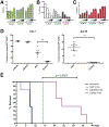
Figure 5.. CB-839 cooperates with HHT in eliminating primary human AML cells in vitro and in vivo.
A,B,C,D) Primary AML cells from four different patients (626, 111, 115, 041) were seeded at 5 × 10⁵/well and treated with CB-839 and HHT alone or together as indicated for 72 h (triangles: 0, 5, 10, and 20 nM HHT; 0, 5 and 10 nM for 041) and the number of viable cells (based on PI exclusion) was counted by flow cytometry. E) Primary AML cells (AML626) were treated with CB-839 and HHT alone or together as in A for 24 h and then added to human methylcellulose complete media. After 16 days, the number of colony forming units was counted. Non-overlapping confidence intervals indicate statistical significance. F) NSG mice were engrafted with primary human AML cells and groups of mice were treated with vehicle (n=5), CB-839 (n=5; 200 mg/kg twice daily p.o.), HHT (n=5; 1 mg/kg once daily i.p.), or CB-839 and HHT in combination (n=10) for 25 days. Leukemic burden was monitored by peripheral blood (PB) draws and quantitation of leukemic cells (human CD45+, HLA-ABC+ cells) by flow cytometry on the indicated days is shown.
Figure 6.
Figure 6.. CB-839 cooperates with HHT in eliminating ALL cells in vitro and in vivo.
A, B, C) Z-119 B-ALL cells were treated with CB-839 and HHT alone or together as indicated for 36 h (triangles: 0, 5, 10, and 20 nM HHT) and mitoROS were measured using MitoPY1 (A). After 72h, the number of viable cells (based on PI exclusion) was counted by flow cytometry (B) and apoptosis was measured after 48 h (C). D) ARF−/− p185 Bcr-Abl/GFP mouse B-ALL cells were transplanted into Bl6 mice and after 5 days, treatment was initiated with vehicle (n=5), CB-839 (n=5; 200 mg/kg twice daily p.o.), HHT (n=5; 1 mg/kg once daily i.p.), or CB-839 and HHT in combination (n=5) for up to 15 days (5 days on/2 days off therapy). After 7 and 10 days of therapy, peripheral blood from all mice was immunostained for B220 and Mac-1 and analyzed by flow cytometry. The percentage of GFP+ cells in the B-lineage (B220+, Mac-1) population was determined and plotted. E) Kaplan-Meier curve showing survival of mice receiving the indicated therapy. Mice were sacrificed when moribund, and all showed clear evidence of leukemia in blood, bone marrow and spleen. For A and C, asterisks indicate statistical significance for comparisons of CB-839 treated cells with control cells (0 CB) under the same HHT treatment conditions. For E, statistical significance determined using the log-rank (Mantel-Cox) test.

References

    1. De Kouchkovsky I, Abdul-Hay M. ‘Acute myeloid leukemia: a comprehensive review and 2016 update’. Blood cancer journal 2016;6:e441. - PMC - PubMed
    1. Kayser S, Levis MJ. Advances in targeted therapy for acute myeloid leukaemia. Br J Haematol 2018;180:484–500 - PMC - PubMed
    1. Bose P, Vachhani P, Cortes JE. Treatment of Relapsed/Refractory Acute Myeloid Leukemia. Current treatment options in oncology 2017;18:17. - PubMed
    1. Leick MB, Levis MJ. The Future of Targeting FLT3 Activation in AML. Current hematologic malignancy reports 2017 - PubMed
    1. Cantor JR, Sabatini DM. Cancer cell metabolism: one hallmark, many faces. Cancer discovery 2012;2:881–98 - PMC - PubMed

Publication types

MeSH terms