. 2019 May 15;79(10):2536-2548.
doi: 10.1158/0008-5472.CAN-18-2170. Epub 2019 Apr 2.

Temozolomide Treatment Induces lncRNA MALAT1 in an NF-κB and p53 Codependent Manner in Glioblastoma

Affiliations

Temozolomide Treatment Induces lncRNA MALAT1 in an NF-κB and p53 Codependent Manner in Glioblastoma

David J Voce et al. Cancer Res. .

Abstract

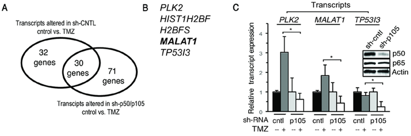
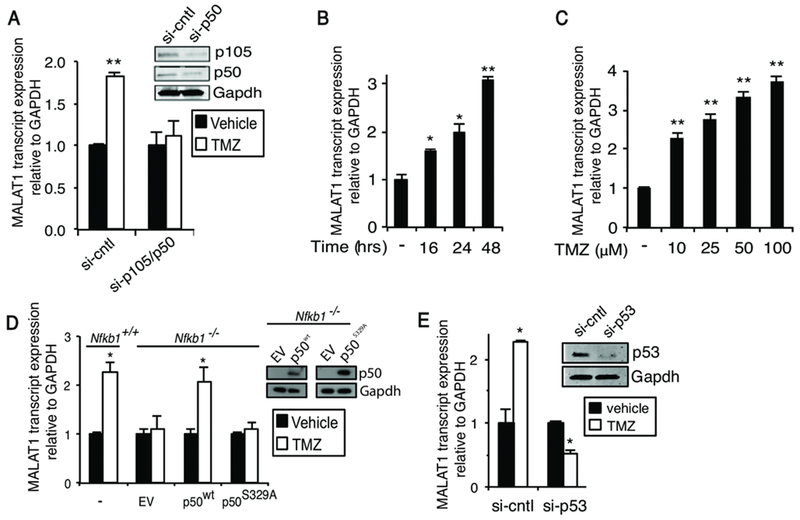
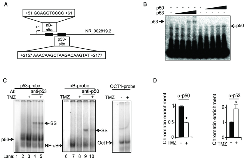
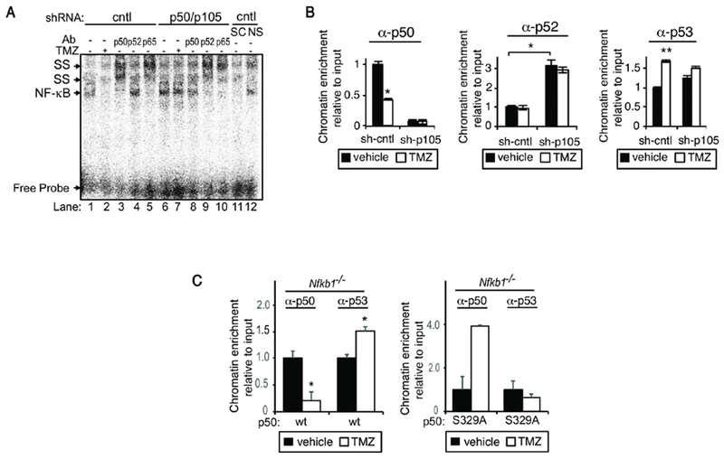
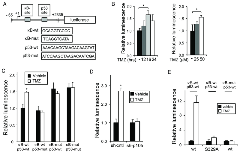
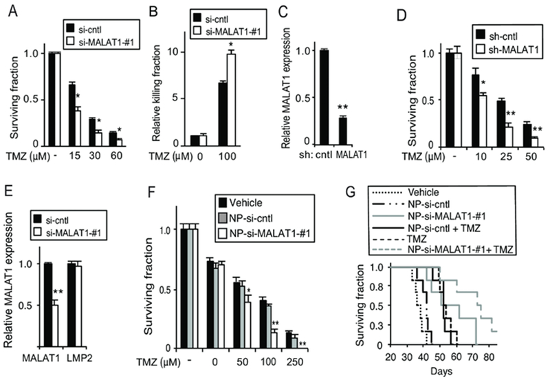
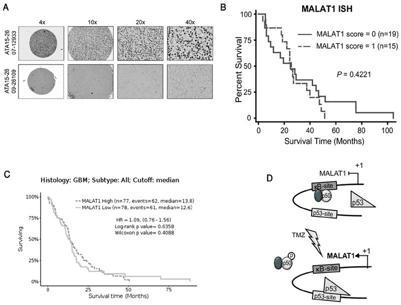
Alkylating chemotherapy is a central component of the management of glioblastoma (GBM). Among the factors that regulate the response to alkylation damage, NF-κB acts to both promote and block cytotoxicity. In this study, we used genome-wide expression analysis in U87 GBM to identify NF-κB-dependent factors altered in response to temozolomide and found the long noncoding RNA (lncRNA) MALAT1 as one of the most significantly upregulated. In addition, we demonstrated that MALAT1 expression was coregulated by p50 (p105) and p53 via novel κB- and p53-binding sites in the proximal MALAT1 coding region. Temozolomide treatment inhibited p50 recruitment to its cognate element as a function of Ser329 phosphorylation while concomitantly increasing p53 recruitment. Moreover, luciferase reporter studies demonstrated that both κB and p53 cis-elements were required for efficient transactivation in response to temozolomide. Depletion of MALAT1 sensitized patient-derived GBM cells to temozolomide cytotoxicity, and in vivo delivery of nanoparticle-encapsulated anti-MALAT1 siRNA increased the efficacy of temozolomide in mice bearing intracranial GBM xenografts. Despite these observations, in situ hybridization of GBM specimens and analysis of publicly available datasets revealed that MALAT1 expression within GBM tissue was not prognostic of overall survival. Together, these findings support MALAT1 as a target for chemosensitization of GBM and identify p50 and p52 as primary regulators of this ncRNA. SIGNIFICANCE: These findings identify NF-κB and p53 as regulators of the lncRNA MALAT1 and suggest MALAT1 as a potential target for the chemosensitization of GBM.


Conflict of interest statement

Conflict of Interest: LNK Chemsolutions, LLC (R.S., L.N. and G.F.L.) have commercial interests in the nanoparticles described.

Figures

Figure 1.
Figure 1.. Gene expression analysis.
(A) Number of transcripts significantly altered in response to TMZ (100 μM, 16 hrs) in U87 cells expressing indicated shRNA (FDR<0.05). (B) List of genes most significantly altered (FDR < 0.01) following TMZ treatment in cells expressing sh-control but not sh-p50/p105. (C) RNA expression in U87 cells expressing sh-control or sh-p105 following treatment with TMZ (100 μM, 16 hrs). Data show mean expression relative to GAPDH, ±SD of triplicate samples from two biological experiments normalized to vehicle. Inset: immunoblot in U87 cells expressing control or p105 shRNA. *, p< 0.05.
Figure 2.
Figure 2.. MALAT1 is induced in a p50/p53-depedent manner.
qPCR analysis of MALAT1 expression. (A) U87 cells transfected with the indicated si-RNA (si-cntl: scrambled sequence) and treated as shown (100 μM TMZ, 24 hours). Inset: immunoblot in U87 cells expressing control or p105 si-RNA. (B) U87 cells treated with 100 μM TMZ for the indicated time. (C) U87 cells treated for 48 hours with TMZ. (D) Nfkb1+/+ and Nfkb1−/− MEFs untransfected or transfected with empty vector (EV), p50wt or p50S329A and treated as indicated (TMZ, 100 μM, 24 hours). Inset: immunoblot in Nfkb1−/− MEFs expressing EV, S329A or wt-p50. (E) U87 cells transfected with si-RNA against p53 and treated as shown (100 μM TMZ, 24 hours). Inset: immunoblot in U87 cells transected with p53 si-RNA. Data show mean expression relative to GAPDH, ±SD of triplicate samples from three biological experiments normalized to vehicle. *, p< 0.05; **, p< 0.01.
Figure 3.
Figure 3.. TMZ modulates binding of p53 and NF-кB to кB and p53 binding sites.
(A) Schematic representation of the кB and p53 binding sites in human MALAT1. (B) EMSA using the кB and p53 probes with increasing concentrations of purified His-p50 or p53 (0 - 300 ng). (C) EMSA using nuclear extracts from U87 cells treated with vehicle or TMZ (100 μM TMZ, 16 hours). Supershift (SS) with anti-p50 and anti-p53. Oct1 EMSA demonstrates equal lysate loading. (D) qChIP using primers spanning the кB and p53 BSs in U87 cells treated as shown (100 μM TMZ, 16 hours). Data represent chromatin enrichment of the indicated protein, relative to input DNA after controlling for non-specific binding using anti-histone H1 (positive control) and anti-IgG, normalized to vehicle, mean ±SD of triplicate samples shown. *, p< 0.05.
Figure 4.
Figure 4.. Recruitment to the кB and p53 binding sites is dependent on p105/p50.
(A) EMSA using nuclear extract from U87 cells stably expressing sh-p105/p50 or sh-control treated with vehicle or TMZ (100 μM, 16 hours). SS with the indicated antibody. Competition performed with specific (SC) and non-specific (NS) cold probe identifies the NF-кB band. (B) qChIP using primers spanning the human кB and p53 BSs in sh-cntl and sh-p105/p50 U87 cells. Cells were treated as shown (100 μM TMZ, 16 hours) and IP performed with the indicated antibodies. (C) qChIP in Nfkb1−/− MEFs expressing either p50wt or p50S329A using primers spanning murine BSs in MALAT1. qChIP data represent chromatin enrichment of the indicated protein, relative to input DNA after controlling for non-specific binding using anti-histone H3 (positive control) and anti-IgG, normalized to vehicle, mean ± SD of triplicate samples, repeated with similar results. *, p< 0.05; **, p< 0.01.
Figure 5.
Figure 5.. MALAT1 кB and p53 binding sites are functional.
(A) Schematic of the 2.4 kbp luciferase reporter containing putative human p53 and p50 BSs. (B-E) Luciferase assays. Data show mean luciferase expression relative to renilla, ±SD of triplicate samples. (B) U87 cells treated with 100 μM TMZ for different times as noted (left) or U87 cells treated with TMZ for 16 hours (right). (C) U87 cells transfected with the indicated reporter construct and treated as shown (100 μM TMZ, 16 hours). (D) sh-cntl and sh-p105/p50 U87 cells treated as in C. (E) U87 cells stably expressing sh-p105/p50 were transfected with p50wt or p50S329A and the indicated кB reporter (p53-wt). Cells were treated as in C. Data represent mean ±SD of triplicate samples, repeated with similar results. *, p < 0.05, **p < 0.01.
Figure 6.
Figure 6.. Knockdown of MALAT1 enhances the anti-glioma effect of TMZ.
(A) Clonogenic assay in U87 cells transfected with si-cntrl or si-MALAT1-#1 treated as shown. (B) Trypan blue assay in GBM34 GSCs transfected with si-cntrl or si-MALAT1-#1 following treatment with TMZ for 72 hours. (C) MALAT1 expression in U87 cells following infection with the indicated sh-RNA construct. (D) Clonogenic assay in U87 cells expressing sh-control or sh-MALAT1 treated with TMZ. (E) qPCR analysis of MALAT1 and LMP2 mRNA expression, relative to GAPDH, in U87 cells treated with nanoparticles carrying si-control (NP-si-cntrl) or si-MALAT1 (NP-si-Malat1). (F) Clonogenic assay in U87 cells following treatment with the indicated nanoparticles. (G) Kaplan-Meier survival curves of mice bearing intracranial U87 GBM xenografts (n= 6 mice per group) following treatment with TMZ (days 4, 7 and 10) and/or the indicated NP. P < 0.02, Log-rank: TMZ + NP-si-MALAT1-#1 vs. TMZ + NP-si-cntl or NP-si-MALAT1-#1 alone. Trypan blue, clonogenic, qPCR data represent mean ±SD of triplicate samples, repeated with similar findings. *, p< 0.05; **, p< 0.01 relative to control.
Figure 7.
Figure 7.. MALAT1 expression is not prognostic of overall survival in GBM.
(A) Representative MALAT1 in-situ hybridization staining in GBM. Low staining (bottom) and high staining (top). (B) Kaplan-Meier survival curves in thirty-four GBM patients separated by their MALAT1 ISH staining score. Significance was analyzed by Log-rank method. (C) Kaplan-Meier survival curves in all GBM patients from TCGA based on RNA-SEQ expression separated at median value. (D) Model illustrating the mechanism by which TMZ induces MALAT1 expression. p50-containing dimers occupy the кB-site at rest (upper). Following treatment (lower), phosphorylation of p50 Ser329 leads to decreased p50 dimer binding and a concomitant increased p53 chromatin recruitment resulting in increased MALAT1 expression.

References

    1. Hegi ME, Diserens A-C, Gorlia T, Hamou M-F, de Tribolet N, Weller M, et al. MGMT gene silencing and benefit from temozolomide in glioblastoma. N Engl J Med [Internet]. 2005;352:997–1003. - PubMed
    1. Perkins ND. The diverse and complex roles of NF-кB subunits in cancer. Nat Rev Cancer. 2012;12:121–32. - PubMed
    1. Schmitt AM, Crawley CD, Kang S, Raleigh DR, Yu X, Wahlstrom JS, et al. p50 (NF-kappaB1) is an effector protein in the cytotoxic response to DNA methylation damage. Mol Cell [Internet]. 2011;44:785–96. - PMC - PubMed
    1. Raychaudhuri B, Han Y, Lu T, Vogelbaum MA. Aberrant constitutive activation of nuclear factor kappaB in glioblastoma multiforme drives invasive phenotype. J Neurooncol. 2007;85:39–47. - PubMed
    1. Yamini B, Yu X, Dolan ME, Wu MH, Darga TE, Kufe DW, et al. Inhibition of nuclear factor-kappaB activity by temozolomide involves O6-methylguanine induced inhibition of p65 DNA binding. Cancer Res [Internet]. 2007;67:6889–98. - PubMed

Publication types