. 2019 Apr 2;10(2):e00014-19.
doi: 10.1128/mBio.00014-19.

Identification of a Novel Deltavirus in Boa Constrictors

Affiliations

Identification of a Novel Deltavirus in Boa Constrictors

Udo Hetzel et al. mBio. .

Abstract

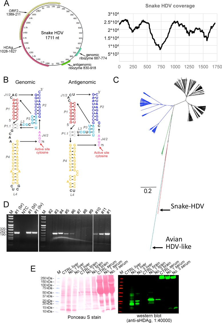
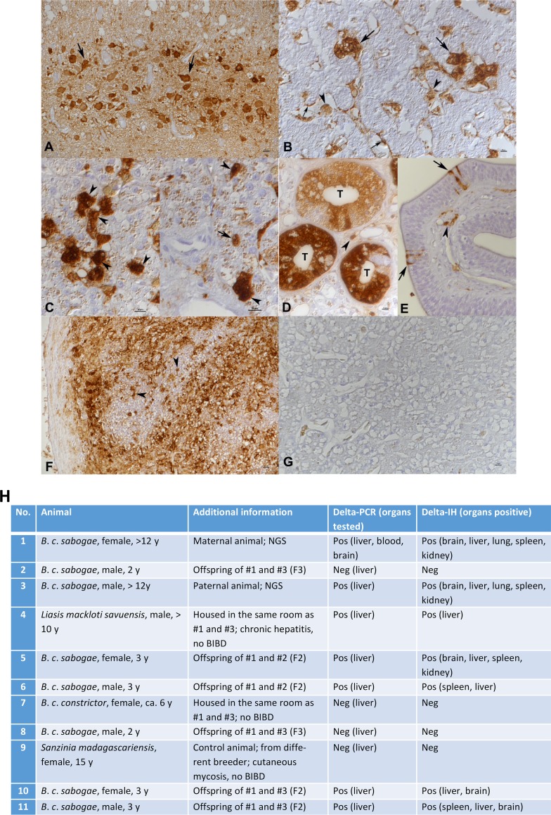
Hepatitis D virus (HDV) forms the genus Deltavirus unassigned to any virus family. HDV is a satellite virus and needs hepatitis B virus (HBV) to make infectious particles. Deltaviruses are thought to have evolved in humans, since for a long time, they had not been identified elsewhere. Herein we report, prompted by the recent discovery of an HDV-like agent in birds, the identification of a deltavirus in snakes (Boa constrictor) designated snake HDV (sHDV). The circular 1,711-nt RNA genome of sHDV resembles human HDV (hHDV) in its coding strategy and size. We discovered sHDV during a metatranscriptomic study of brain samples of a Boa constrictor breeding pair with central nervous system signs. Applying next-generation sequencing (NGS) to brain, blood, and liver samples from both snakes, we did not find reads matching hepadnaviruses. Sequence comparison showed the snake delta antigen (sHDAg) to be 55% and 37% identical to its human and avian counterparts. Antiserum raised against recombinant sHDAg was used in immunohistology and demonstrated a broad viral target cell spectrum, including neurons, epithelial cells, and leukocytes. Using RT-PCR, we also detected sHDV RNA in two juvenile offspring and in a water python (Liasis macklotisavuensis) in the same snake colony, potentially indicating vertical and horizontal transmission. Screening of 20 randomly selected boas from another breeder by RT-PCR revealed sHDV infection in three additional snakes. The observed broad tissue tropism and the failure to detect accompanying hepadnavirus suggest that sHDV could be a satellite virus of a currently unknown enveloped virus.IMPORTANCE So far, the only known example of deltaviruses is the hepatitis delta virus (HDV). HDV is speculated to have evolved in humans, since deltaviruses were until very recently found only in humans. Using a metatranscriptomic sequencing approach, we found a circular RNA, which resembles that of HDV in size and coding strategy, in a snake. The identification of similar deltaviruses in distantly related species other than humans indicates that the previously suggested hypotheses on the origins of deltaviruses need to be updated. It is still possible that the ancestor of deltaviruses emerged from cellular RNAs; however, it likely would have happened much earlier in evolution than previously thought. These findings open up completely new avenues in evolution and pathogenesis studies of deltaviruses.

Keywords: deltavirus; evolutionary biology; hepatitis; virology; zoonotic infections.


Figures

FIG 1
FIG 1
Genome organization, sequencing coverage, schematic ribozyme structure, and phylogenetic analysis of snake HDV. (A) Schematic presentation of circular RNA genome and sequencing coverage for snake HDV. The genome shows two open reading frames (ORFs). The ORF in the antigenomic orientation spanning nucleotide residues 1028 to 1627 encodes a 199-amino-acid protein which by BLAST analysis represents the HDAg. ORF2 is in the genomic orientation and spans residues 1389 to 211 and encodes a 177-amino-acid protein, which by BLAST analysis did not yield significant hits (35% identity over 66 amino acids (E value of 5) to ferritin-like protein from “Candidatus Nitrososphaera evergladensis” SR1, NCBI protein accession no. AIF82718.1). SMART (Simple Modular Architecture Research Tool available at http://smart.embl-heidelberg.de/) analysis showed the putative protein to have two transmembrane helices, and a DUF3343 (domain of unknown function) domain with an E value of 0.013. The genomic and antigenomic ribozymes identified by sequence alignments to known HDVs are located at nt 687 to 744 and 830 to 918, respectively. The graph shows sequencing coverage (on the y axis) in respect to each nucleotide position (on the x axis) of snake HDV from the original brain sample, and coverage ranges from 7,368 (at nt position 729) to 26,304-fold. (B) Models for the secondary structures of the genomic and antigenomic ribozymes identified in snake HDV. The presentation format is adopted from a review by Webb and Luptak (20) which was also used by Wille et al. (2). Paired regions (P), joining regions (J), and loops (L) are shown. Both genomic and antigenomic ribozymes are structurally close to their human HDV counterparts described in reference , and they are identical at the following regions: active site, P1.1, and P3. Cleavage by the ribozyme occurs at the 5′ end. (C) Phylogenetic analysis of human, avian, and reptile HDAgs. The phylogenetic analysis was done using Bayesian MCMC method implemented in MrBayes 3.1.2 (21) with the JTT model of substitution with gamma distributed rate variation among sites. HDV genotype 1 (black), HDV genotype 2 (blue), HDV genotype 3 (green), avian HDV-like sequence (cyan), and snake HDV (red) are indicated. (D) RT-PCR results of snake tissues. The gel on the left shows RT-PCR products obtained for snake 1 (Fig. 2H) from different tissues: brain (br), blood (bl), and liver (liv). NTC, nontemplate control, M is DNA ladder. The gel on the right shows RT-PCR products obtained from liver samples, the animal numbering is according to Fig. 2H, and animal 1 serves as a positive control. (E) Western blot of liver and brain homogenates and serum from sHDV RT-PCR negative-control animal (animal 7 [Fig. 2H]) and sHDV RT-PCR-positive animals (animals 1 and 3 [Fig. 2H]). The panel on the left shows total protein staining by Ponceau S, and the panel on the right shows staining with anti-sHDAg (1:40,000) antiserum using IRDye 800CW-conjugated donkey anti-rabbit IgG (LI-COR Biosciences). The signal for Western blot was read with Odyssey Infrared Imaging System (LI-COR Biosciences).
FIG 2
FIG 2
Immunohistology for sHDAg in NGS- and RT-PCR-positive (animals 1 and 3 [A to F]) and negative (animal 7 [G]) animals, and a table of animals included in the study. (A) Brain. Viral antigen is expressed in the nucleus, cytoplasm, and cell processes of numerous neurons. (B) Liver. Individual hepatocytes (large arrows) are strongly positive, and macrophages (arrowheads) and endothelial cells (small arrows) are found to also express viral antigen. (C) Liver. A closer view shows that a substantial proportion of hepatocytes exhibit both cytoplasmic and nuclear (arrowheads) sHDAg expression. On the right, there is also one individual hepatocyte with an exclusively nuclear reaction (arrow). (D) Kidney. In a group of tubules (T), the majority of epithelial cells exhibit variably intense viral antigen expression. Occasional leukocytes in the interstitium (arrowhead) are also positive. (E) Lung. There are several individual positive epithelial cells (arrows); some subepithelial leukocytes are also found to express viral antigen (arrowhead). (F) Spleen. There is extensive viral antigen expression. Positive cells often have the morphology of macrophages (arrowheads). (G) RT-PCR-negative animal (animal 7), liver immunohistology for sHDAg. There is no evidence of sHDAg expression. Horseradish peroxidase method, hematoxylin counterstain. Note that the finely granular brownish staining in some Kupffer cells and hepatocytes in panels C and G is due to bile pigment and/or hemosiderin. (H) Table of animals included in the study.

References

    1. Le Gal F, Gault E, Ripault MP, Serpaggi J, Trinchet JC, Gordien E, Deny P. 2006. Eighth major clade for hepatitis delta virus. Emerg Infect Dis 12:1447–1450. doi:10.3201/eid1209.060112. - DOI - PMC - PubMed
    1. Wille M, Netter HJ, Littlejohn M, Yuen L, Shi M, Eden J, Klaassen M, Holmes EC, Hurt AC. 2018. A divergent hepatitis D-like agent in birds. bioRxiv doi:10.1101/423707. - DOI - PMC - PubMed
    1. Lempp FA, Ni Y, Urban S. 2016. Hepatitis delta virus: insights into a peculiar pathogen and novel treatment options. Nat Rev Gastroenterol Hepatol 13:580–589. doi:10.1038/nrgastro.2016.126. - DOI - PubMed
    1. Littlejohn M, Locarnini S, Yuen L. 2016. Origins and evolution of hepatitis B virus and hepatitis D virus. Cold Spring Harb Perspect Med 6:a021360. doi:10.1101/cshperspect.a021360. - DOI - PMC - PubMed
    1. Wang KS, Choo QL, Weiner AJ, Ou JH, Najarian RC, Thayer RM, Mullenbach GT, Denniston KJ, Gerin JL, Houghton M. 1986. Structure, sequence and expression of the hepatitis delta (delta) viral genome. Nature 323:508–514. doi:10.1038/323508a0. - DOI - PubMed

Publication types

MeSH terms