Review
. 2019 May;20(5):357-370.
doi: 10.1038/s41435-019-0065-0. Epub 2019 Apr 3.

Yersinia pestis and plague: an updated view on evolution, virulence determinants, immune subversion, vaccination, and diagnostics

Affiliations
Review

Yersinia pestis and plague: an updated view on evolution, virulence determinants, immune subversion, vaccination, and diagnostics

Christian E Demeure et al. Genes Immun. 2019 May.

Abstract

Plague is a vector-borne disease caused by Yersinia pestis. Transmitted by fleas from rodent reservoirs, Y. pestis emerged <6000 years ago from an enteric bacterial ancestor through events of gene gain and genome reduction. It is a highly remarkable model for the understanding of pathogenic bacteria evolution, and a major concern for public health as highlighted by recent human outbreaks. A complex set of virulence determinants, including the Yersinia outer-membrane proteins (Yops), the broad-range protease Pla, pathogen-associated molecular patterns (PAMPs), and iron capture systems play critical roles in the molecular strategies that Y. pestis employs to subvert the human immune system, allowing unrestricted bacterial replication in lymph nodes (bubonic plague) and in lungs (pneumonic plague). Some of these immunogenic proteins as well as the capsular antigen F1 are exploited for diagnostic purposes, which are critical in the context of the rapid onset of death in the absence of antibiotic treatment (less than a week for bubonic plague and <48 h for pneumonic plague). Here, we review recent research advances on Y. pestis evolution, virulence factor function, bacterial strategies to subvert mammalian innate immune responses, vaccination, and problems associated with pneumonic plague diagnosis.


Conflict of interest statement

The authors declare that they have no conflict of interest.

Figures

Fig. 1
Fig. 1
Yersinia pestis phylogenetic tree. Schematic representation of the Y. pestis genealogy, including previously described phylogenetic branches (0–4) and biovars (ANT: Antiqua, MED: Medievalis, ORI: Orientalis, IN: Intermediate, PE: Pestoides, including Microtus isolates), as well as six novel Pestoides clusters [25]. The length of the branches does not reflect the evolutionary time. Nodes from branch 3, branch 2 and branch 1.IN were collapsed for simplicity. The node indicated as an asterisk marks the polytomy known as the «Big Bang», which gave rise to branches 1–4. The three historically recorded plague pandemics are presented in bold next to their associated phylogenetic lineages. The key gene gain and inactivation events in the evolution of Y. pestis from Y. pseudotuberculosis are displayed. These events include the acquisition of two virulence-associated plasmids (pFra/pMT1 and pPla/pPCP1), the gain of Yersinia murine toxin (ymt) in the pFra/pMT1 plasmid, as well as by the inactivation of the virulence-associated genes pde3 (1promoter mutation; 2inactivating mutation), ureD, rcsA, flhD, and pde2. The aftermath of these molecular changes gave rise to the Y. pestis’ flea-borne transmission and thereby the ability to cause bubonic plague. The acquisition of the I259T mutation in the plasminogen activator (Pla), a key virulence factor responsible for the fulminant lung infection specific of Y. pestis, is also presented in the phylogeny
Fig. 2
Fig. 2
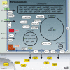
Yersinia pestis virulence determinants. Y. pestis requires the three well-characterized virulence plasmids pYV/pCD1, pPla/pPCP1, and pFra/pMT1, as well as chromosomally encoded virulence factors to cause disease. Examples of cytoplasmic, cell-surface associated, and secreted virulence factors are shown. The infectious process involves adhesion to host cells mediated by Braun lipoproteins Lpp and proteins such as Ail, PsaA, and Pla. Yop effectors, including YopH, YopE, YopT, YopJ, YpkA, YopM, and YopK, are subsequently delivered through the T3SS to trigger apoptosis, inhibit phagocytosis, and block cytokine production. LPS modification and the capsular antigen F1 encoded by the caf gene further contribute to Yersinia immune escape. The expression of many of these virulence determinants is induced during the transition from the temperature of the flea midgut (26 °C) to that of the mammalian host (37 °C). Y. pestis survival in the host requires efficient metal acquisition systems. The yersiniabactin-dependent iron uptake system is encoded in the high-pathogenicity island within the pigmentation chromosomal locus pgm. Other metal transport systems, including YbtX, ZnuABC, Yfe, and Feo, also play a role in infection. Additional virulence factors have been identified by signature-tagged mutagenesis, “per-pool” mutant screening, or in vivo transcriptional profiling, e.g., YMPY1.66c, BrnQ, RbsA, GspE, NirC, CyoABCDE, PspABC, Ypo0862, Ypo1119, Ypo1120, Ypo1501, and Ypo 2884. BCAA: branched-chain amino acids, Fe: iron, LPS: lipopolysaccharide, T2SS: type-two secretion system, T3SS: type-three secretion system, T6SS: type-six secretion system, Ybt: yersiniabactin, Yop: Yersinia outer-membrane protein, Zn: zinc. Components of the illustration are not drawn to scale
Fig. 3
Fig. 3
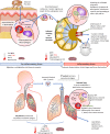
Innate immunity subversion by Yersinia pestis. Schematic representation of Y. pestis transmission and migration in the human body during bubonic (upper panel) and pneumonic plague (lower panel). The effect of temperature of origin and its effects on the bacterium are indicated at various steps. A focus is given on innate immunity host cells encountered by the pathogen and involved in immune evasion mechanisms described in the text, which are shown as zoomed areas
Fig. 4
Fig. 4
Plague diagnostics: from presumption to confirmation. Upon plague suspicion, according to epidemiological and clinical criteria, collected samples (sputum, expectoration) can be analyzed for the detection of the Y. pestis capsular antigen F1 using the rapid diagnostic test (RDT: the C band is a migration control, and the T band reveals the presence of F1). Confirmation is performed through detection of Y. pestis DNA using real-time PCR targeting pla and caf1, and in the case of discordant or uncertain results, a PCR targeting pla, caf1, and inv is performed. Isolation of Y. pestis remains the gold standard in biological diagnostic tests: culture on selective CIN media is followed by bacterial identification using biochemical tests or mass spectrometry, confirmation using PCR, and phage lysis. Boxed area: algorithm for molecular tests using qPCR and PCR (reprinted from ref. [135])

References

    1. Stenseth NC, Atshabar BB, Begon M, Belmain SR, Bertherat E, Carniel E, et al. Plague: past, present, and future. PLoS Med. 2008;5:e3. - PMC - PubMed
    1. Perry RD, Fetherston JD. Yersinia pestis—etiologic agent of plague. Clin Microbiol Rev. 1997;10:35–66. - PMC - PubMed
    1. Yersin A. La peste bubonique a Hong-Kong. Ann Inst Pasteur (Paris) 1894;8:662–7.
    1. Vogler AJ, Chan F, Nottingham R, Andersen G, Drees K, Beckstrom-Sternberg SM, et al. A decade of plague in mahajanga, madagascar: insights into the global maritime spread of pandemic plague. mBio. 2013;4:e00623–12–e00623–12. - PMC - PubMed
    1. Bertherat E. Plague around the world, 2010–2015. Wkly Epidemiol Rec. 2016;91:89–104.

Publication types

Substances