

Polysaccharides Cell Wall Architecture of Mucorales
- PMID: 30941108
- PMCID: PMC6433966
- DOI: 10.3389/fmicb.2019.00469
Polysaccharides Cell Wall Architecture of Mucorales
Abstract
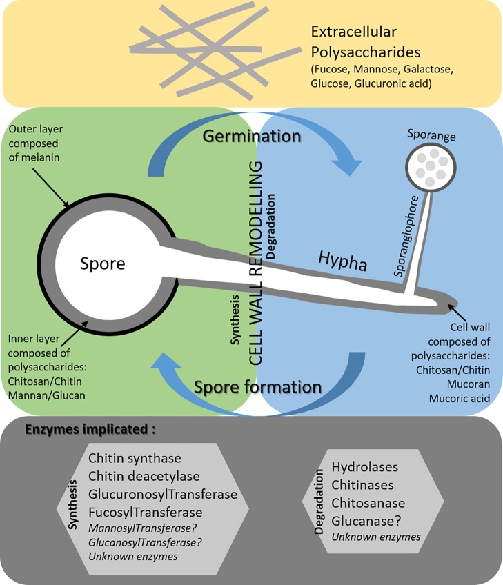
Invasive fungal infections are some of the most life-threatening infectious diseases in the hospital setting. In industrialized countries, the most common fungal species isolated from immunocompromised patients are Candida and Aspergillus spp. However, the number of infections due to Mucorales spp. is constantly increasing and little is known about the virulence factors of these fungi. The fungal cell wall is an important structure protecting fungi from the environment. A better knowledge of its composition should improve our understanding of host-pathogen interactions. Cell wall molecules are involved in tissue adherence, immune escape strategies, and stimulation of host defenses including phagocytosis and mediators of humoral immunity. The fungal cell wall is also a target of choice for the development of diagnostic or therapeutic tools. The present review discusses our current knowledge on the cell wall structure of Mucorales in terms of the polysaccharides and glyco-enzymes involved in its biosynthesis and degradation, with an emphasis on the missing gaps in our knowledge.
Keywords: Mucorales; cell wall; glucuronic acid; glyco-enzymes; polysaccharides.
Figures
References
-
- Alfonso C., Jesús Martínez M., Reyes F. (1992). Purification and properties of two endochitosanases from Mucor rouxii implicated in its cell wall degradation. FEMS Microbiol. Lett. 95, 187–194. 10.1111/j.1574-6968.1992.tb05364.x - DOI
-
- Almeida C. A., de Camos-Takaki G. M., Portela M. B., Travassos L. R., Alviano C. S., Alviano D. S. (2013). Sialoglycoproteins in morphological distinct stages of Mucor polymorphosphorus and their influence on phagocytosis by human blood phagocytes. Mycopathologia 176, 183–189. 10.1007/s11046-013-9692-6, PMID: - DOI - PubMed
Publication types
LinkOut - more resources
Full Text Sources
Miscellaneous