Review
. 2019 Mar 19:10:504.
doi: 10.3389/fimmu.2019.00504. eCollection 2019.

The Emerging Role of Complement Proteins as a Target for Therapy of IgA Nephropathy

Affiliations
Review

The Emerging Role of Complement Proteins as a Target for Therapy of IgA Nephropathy

Dana V Rizk et al. Front Immunol. .

Abstract

IgA nephropathy (IgAN) is the most common form of primary glomerulonephritis worldwide and a common cause of end-stage renal disease. Evaluation of a kidney biopsy is necessary for diagnosis, with routine immunofluorescence microscopy revealing dominant or co-dominant IgA immunodeposits usually with complement C3 and sometimes IgG and/or IgM. IgA nephropathy reduces life expectancy by more than 10 years and leads to kidney failure in 20-40% of patients within 20 years of diagnosis. There is accumulating clinical, genetic, and biochemical evidence that complement plays an important role in the pathogenesis of IgA nephropathy. The presence of C3 differentiates the diagnosis of IgA nephropathy from the subclinical deposition of glomerular IgA. Markers for the activation of the alternative and mannan-binding lectin (MBL) pathways in renal-biopsy specimens are associated with disease activity and portend a worse renal outcome. Complement proteins in the circulation have also been evaluated in IgA nephropathy and found to be of prognostic value. Recently, genetic studies have identified IgA nephropathy-associated loci. Within these loci are genes encoding products involved in complement regulation and interaction with immune complexes. Put together, these data identify the complement cascade as a rational treatment target for this chronic kidney disease. Recent case reports on the successful use of humanized anti-C5 monoclonal antibody eculizumab are consistent with this hypothesis, but a better understanding of the role of complement in IgA nephropathy is needed to guide future therapeutic interventions.

Keywords: IgA nephropathy; IgAN; IgAN pathogenesis; IgAN treatment; alternative complement pathway; complement; mannan binding lectin complement pathway.


Figures

Figure 1
Figure 1
Proposed four-hit pathogenesis of IgAN. Circulatory galactose-deficient IgA1 (Gd-IgA1) (Hit 1) is recognized by specific autoantibodies (Hit 2) to form circulating immune complexes (Hit 3). Some of these immune complexes deposit in the kidneys, thereby leading to mesangial activation, enhanced proliferation of mesangial cells, and ultimately kidney injury (Hit 4). Certain genetic loci have been associated with increased risk for developing IgAN. The activation of the alternative and, at least in some patients, mannan-binding lectin (MBL) pathways by immune complexes is involved in disease pathogenesis.
Figure 2
Figure 2
Complement activation pathways and examples of complement-targeting therapeutics for IgAN. The three pathways of complement activation, classical, lectin, and alternative, are initiated by interactions of complement proteins with distinct structures. Complexes of antigen and antibody can activate the classical pathway. Mannan-binding lectin recognizes carbohydrate structures and, upon association with serine proteases (MASP, mannose-associated serine proteases), can activate the lectin pathway. Complement C3 that is covalently bound to microorganism surfaces as C3b initiates the cascade of the alternative pathway. Each pathway can ultimately generate an active C3 convertase, resulting in cleavage of C3 component into C3a and C3b fragments. C3b can interact with C4b2b or C3bBb to produce C5 convertase that cleaves C5 into C5a and C5b fragments. C5b binds to the cell membrane and serves as a platform for assembly of the membrane attack complex (MAC) (24, 25). The formation of MAC can be inhibited by membrane-bound CD59 that binds to C8 and/or C9. Several other regulatory proteins of the complement-activation pathways are shown in red. Five complement-targeting therapeutics are shown in blue rounded rectangles. These reagents include two monoclonal antibodies (eculizumab that blocks cleavage of C5; OMS721 targeting MASP-2 that inhibits its protease activity), a C5a receptor antagonist (CCX168), a low-molecular-weight inhibitor of factor B (LNP023), and an inhibitor of C3 activation (APL-2). CR1, complement receptor 1; CFHR 1-5, complement factor H-related proteins 1-5; DAF, decay-accelerating factor; FB, factor B; FD, factor D; FI, factor I; Gd-IgA1, galactose-deficient IgA1; Gd-IgA1-IC, galactose-deficient IgA1-containing immune complexes; MCP, membrane cofactor protein; P, properdin.
Figure 3
Figure 3
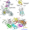
Examples of therapeutic control of complement-activation pathways. (A) The complex of C5 and the Fab derived from eculizumab antibody (PDB ID: 5I5K) (145) are shown in cartoon model with the heavy (H) and light (L) chains of the Fab colored in magenta and blue, respectively. C5 is shaded in pale cyan, while macroglobulin domain 7 (MG7), the site of eculizumab binding on C5, is highlighted in green. C5a is yellow and the C5a-receptor-activating pentapeptide cleavage site is red. (B) C5a (yellow) binds to the membrane-associated C5a receptor (C5aR, gray background) via the C-terminal C5a pentapeptide (red). CCX168 (Avacopan, yellow stick model) binds to the surface of C5aR (pocket shaded in cyan), thereby blocking C5a binding through allosteric effects on the C5a-binding pocket. The C5a/C5aR model is based on PDB ID: 6C1R (146). (C) Compstatin, and APL-2 are inhibitors of activation of C3. The crystal structure of C3c (PDB ID: 2QKI) (147), a major proteolytic fragment of C3, is shown in complex with compstatin (yellow stick model). Both inhibitors bind to a site (shaded in blue) formed by the macroglobulin domains 4 and 5 (MG4, MG5). All illustrations were prepared with PyMOL (148).
Figure 4
Figure 4
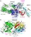
Targets and therapeutic agents of the lectin and alternative complement-activation pathways. (A) The crystal structure of the C4/MASP-2 complex is shown (PDB ID: 4FXG) (149). MASP-2 complement control protein (CCP) domains bind to C4. Upon association with MASP-2, C4 undergoes a conformational change whereby the scissile bond containing R-loop is inserted into the catalytic site of the serine-protease (SP) domain (pale cyan with catalytic triad in magenta). Cleavage yields fragments C4a and C4b. Monoclonal antibody OMS721 binds to a CCP domain of MASP-2, inhibiting the lectin pathway by inhibiting complex formation. (B) The structure of factor B is shown (PDB ID: 2OK5) (150). Upon C3b binding, the N-terminal CCP domains are displaced, leading to rearrangement of the central helices and release of the scissile bond for proteolytic activation. LNP023 inhibits the proteolytic activity of factor B.

References

    1. Berger J, Hinglais N. Intercapillary deposits of IgA-IgG. J Urol Nephrol. (1968) 74:694–5. - PubMed
    1. Berthoux FC, Mohey H, Afiani A. Natural history of primary IgA nephropathy. Semin Nephrol. (2008) 28:4–9. 10.1016/j.semnephrol.2007.10.001 - DOI - PubMed
    1. Hastings MC, Bursac Z, Julian BA, Villa Baca E, Featherston J, Woodford SY, et al. . Life expectancy for patients from the southeastern united states with IgA nephropathy. Kidney Int Rep. (2018) 3:99–104. 10.1016/j.ekir.2017.08.008 - DOI - PMC - PubMed
    1. Jennette JC. The immunohistology of IgA nephropathy. Am J Kidney Dis. (1988) 12:348–52. 10.1016/S0272-6386(88)80022-2 - DOI - PubMed
    1. Conley ME, Cooper MD, Michael AF. Selective deposition of immunoglobulin A1 in immunoglobulin a nephropathy, anaphylactoid purpura nephritis, and systemic lupus erythematosus. J Clin Invest. (1980) 66:1432–6. 10.1172/JCI109998 - DOI - PMC - PubMed

Publication types