Review
. 2019 Jun;31(23):e1900332.
doi: 10.1002/adma.201900332. Epub 2019 Apr 3.

2D Nanoclay for Biomedical Applications: Regenerative Medicine, Therapeutic Delivery, and Additive Manufacturing

Affiliations
Review

2D Nanoclay for Biomedical Applications: Regenerative Medicine, Therapeutic Delivery, and Additive Manufacturing

Akhilesh K Gaharwar et al. Adv Mater. 2019 Jun.

Abstract

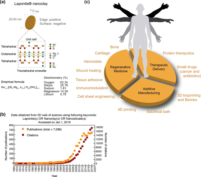
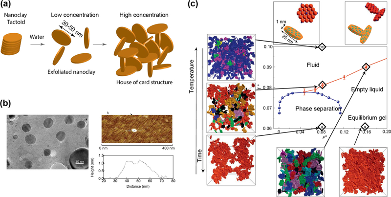
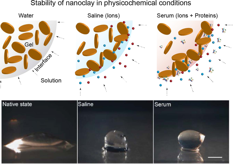
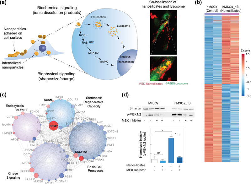
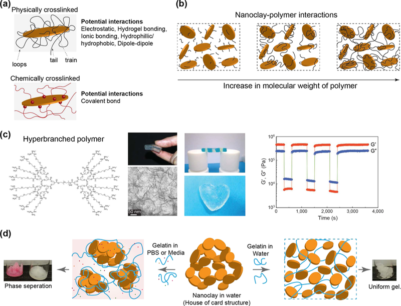
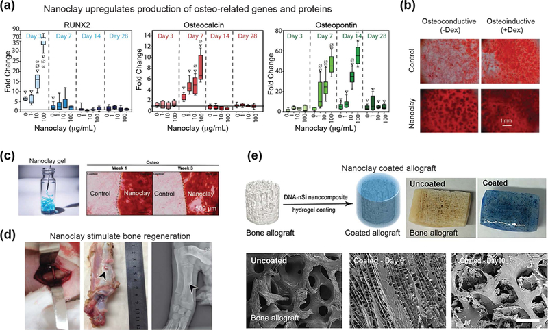
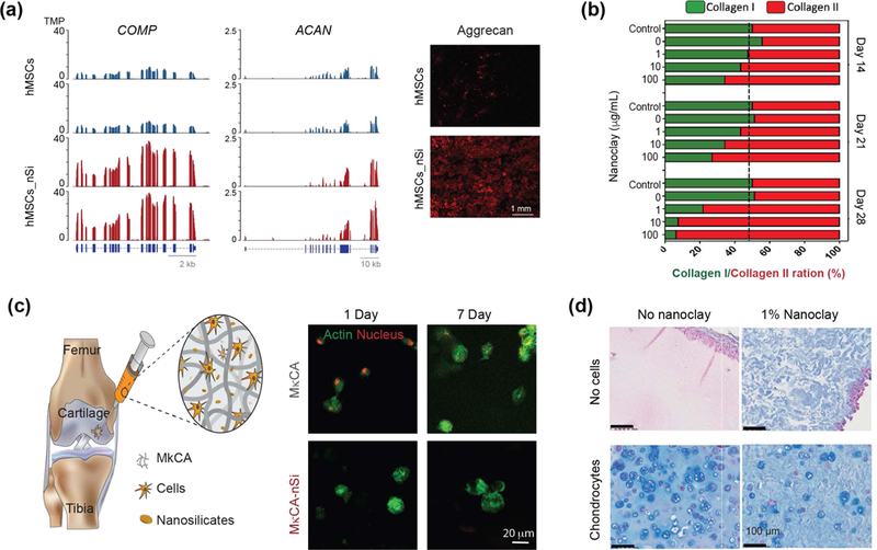
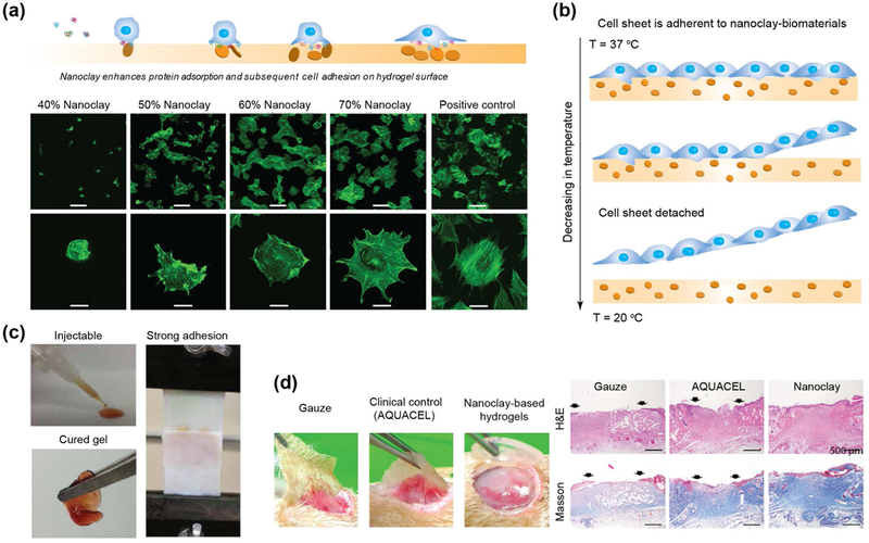
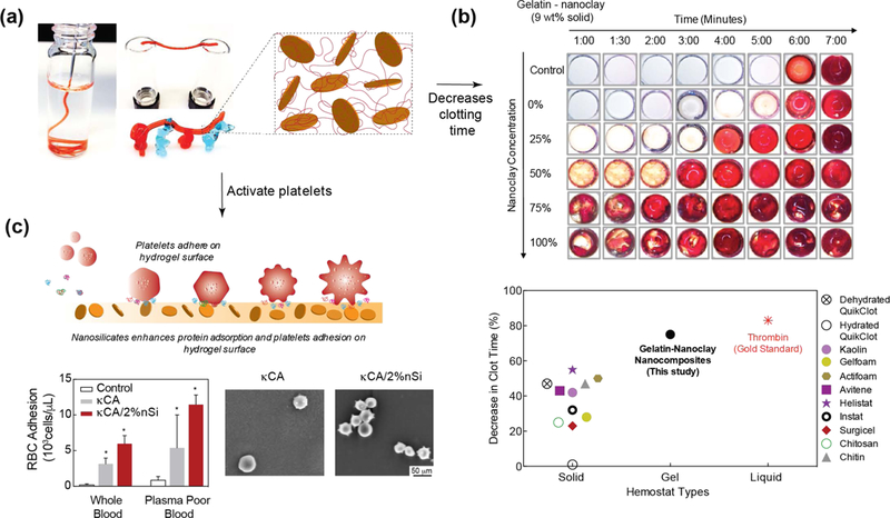
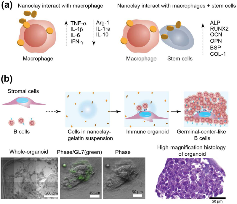
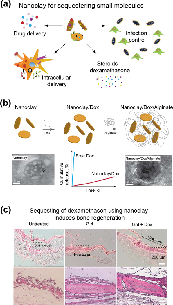
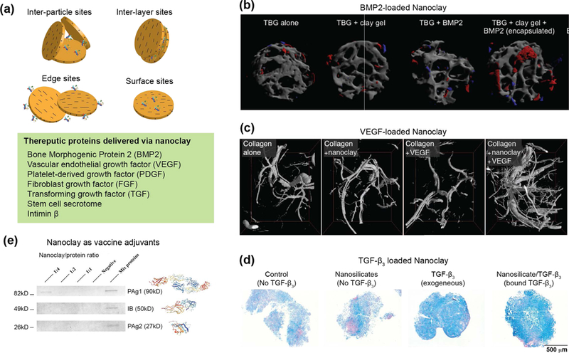
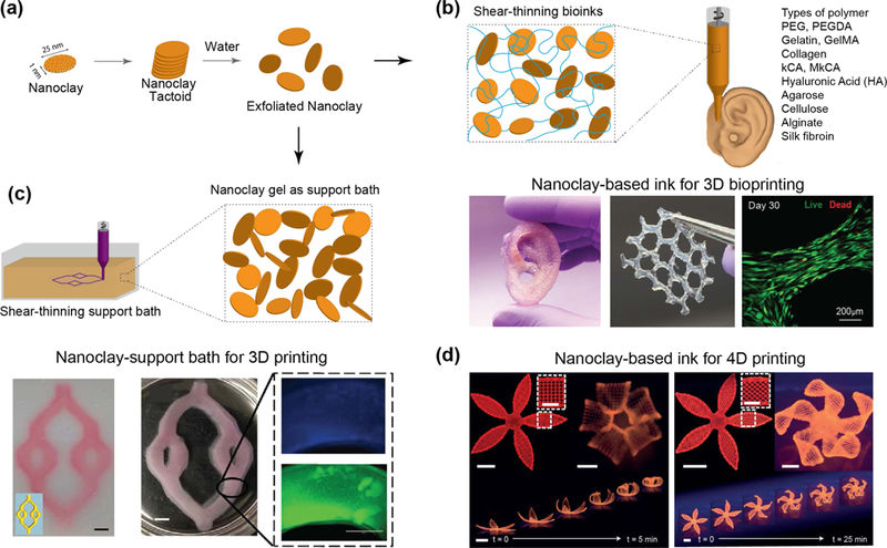
Clay nanomaterials are an emerging class of 2D biomaterials of interest due to their atomically thin layered structure, charged characteristics, and well-defined composition. Synthetic nanoclays are plate-like polyions composed of simple or complex salts of silicic acids with a heterogeneous charge distribution and patchy interactions. Due to their biocompatible characteristics, unique shape, high surface-to-volume ratio, and charge, nanoclays are investigated for various biomedical applications. Here, a critical overview of the physical, chemical, and physiological interactions of nanoclay with biological moieties, including cells, proteins, and polymers, is provided. The state-of-the-art biomedical applications of 2D nanoclay in regenerative medicine, therapeutic delivery, and additive manufacturing are reviewed. In addition, recent developments that are shaping this emerging field are discussed and promising new research directions for 2D nanoclay-based biomaterials are identified.

Keywords: 2D nanomaterials; 3D printing; bioprinting; drug delivery; nanoclay; nanosilicates; tissue engineering.


Figures

Figure 1.
Figure 1.. Nanoclay in biomedical engineering.
(a) Size, shape and chemical composition of individual nanoclay. (b) Exponential increase in investigating nanoclay for various applications. Data obtained from ISI web of science using Laponite(s)/nanoclay(s)/nanosilicates(s) (01 Jan 2019). (c) Biomedical applications of nanoclay are investigated in regenerative medicine, therapeutic delivery and additive manufacturing.
Figure 2.
Figure 2.. Physiochemical characteristics of nanoclay.
(a) Schematic showing exfoliation of nanoclay. At high concentration of nanoclay >3wt%, house-of-card structure is formed that results in formation of gel. (b) Size and shape of individual nanoclay. TEM show circular morphology of individual nanoclay with diameter ~30–50nm, while AFM show thickness of individual nanoclay ~1–2nm. Adapted with permission[13] Copyright © National Academy of Sciences. (c) Phase diagram of nanoclay demonstrate formation of sol-gel transition with respect to concentration, temperature, and time. Adapted with permission[26] Copyright © Springer Nature Publishing AG.
Figure 3.
Figure 3.. Interaction of exfoliated nanoclay with water, saline and serum.
Stability of nanoclay gel in physiological conditions such as water, saline and serum. Adapted and redrawn with permission[95] Copyright © John Wiley & Sons, Inc.
Figure 4.
Figure 4.. Interactions of nanoclay with cells.
(a) Schematic showing interaction of nanoclay with cells. Nanoclay induces both biophysical and biochemical signaling on cells. Co-localization of nanoclay and lysosome within hMSCs (Red – nanoclay; Green – lysosome) determine using microscopy. (b) Normalized gene expression of >4,000 genes following treatment with nanoclay (padjust < 0.05, red (up-regulated): 1,897 genes; blue (down-regulated): 2,171 genes). (c) Gene network of differentially regulated genes after nanoclay treatment. (d) Western blot analysis established role of MAPK/ERK signaling after treatment with nanoclay and MEK inhibitor. Adapted and modified with permission[13] Copyright © National Academy of Sciences.
Figure 5.
Figure 5.. Interactions of nanoclay with polymers.
(a) Nanoclay can form both physical and chemical bonds with polymer chains via multiple mechanisms. (b) Nanoclay interactions with polymer with different molecular weight. Critical size of polymer chain result in formation of physically crosslinked network. (c) Formation of self-assembled gel when nanoclay is mixed with polymeric binder (hyperbranched polymer). Polymer end group interact with nanoclay and result in formation of mechanically stable hydrogels. Adapted with permission[71] Copyright © Springer Nature Publishing AG. (d) Effect of ions on the stability of hydrogels. Adapted and modified with permission[92] Copyright © Royal Society of Chemistry.
Figure 6.
Figure 6.. Osteoinductive characteristics of nanoclay.
(a) Nanoclay treatment in human adipose derived stem cells (hASCs) results in dose-dependent increase in osteo-related gene expression (RUNX2, osteocalcin, and osteopontin). Adapted with permission[55] Copyright © Elsevier. (b) Human mesenchymal stem cells (hMSCs) treated with increasing concentrations of nanoclay produced mineralized matrix in osteoconductive and osteoinductive media. Adapted with permission[54] Copyright © John Wiley & Sons, Inc. (c) Nanoclay gel support adhesion of stem cells and induce osteogenic differentiation as determined by alizarin red staining. Adapted with permission[95] Copyright © John Wiley & Sons, Inc. (d) In vivo bone regeneration is demonstrated by using nanoclay scaffolds. Adapted with permission[96] PLOS ONE. (e) Allograft can be coated with nanoclay-based gels to promote bone formation. Adapted with permission[106] Copyright © American Chemical Society.
Figure 7.
Figure 7.. Nanoclay promote formation of cartilage-related proteins.
(a) Significant upregulation of cartilage related genes such as COMP and ACAN are observed after nanoclay treatment of hMSCs. After 21 days, significant production of ACAN and GAGs are observed from western blot. Adapted with permission[13] Copyright © National Academy of Sciences. (b) Stem cells treated with nanoclay results in enhanced production of collagen II, which is important component of cartilaginous ECM. Adapted with permission[55] Copyright © Elsevier. (c) Nanoclay loaded hydrogels are used for delivery of stem cells for cartilage regeneration. Encapsulated cells show high viability and assume round shape morphology after 7 days indicating potential application in cartilage regeneration. Adapted and modified with permission[114] Copyright © Royal Society of Chemistry. (d) Nanoclay loaded in silated hydroxypropylmethyl cellulose showed enhanced production of cartilaginous ECM as demonstrated by Alcian blue straining by encapsulated chondrocytes. Adapted with permission[117] Copyright © Elsevier.
Figure 8.
Figure 8.. Nanoclay support cell and tissue adhesion.
(a) The addition of nanoclay to a non-fouling polymer (PEO) results in enhanced cell adhesion and spreading. Adapted and modified with permission [98] Copyright © Elsevier. (b) Concept of cell sheet engineering using nanoclay and thermoresponsive polymer. (c) Dopamine-modified multi-armed PEG (PEG-D) mixed with nanoclay was developed as tissue adhesive. Injectability combined with wet tissue adhesion allow this fit-to shape sealant to be used as an injectable bandage. Adapted with permission[134] Copyright © American Chemical Society. (d) nanoclay-based hydrogels facilitate complete closure in normal and diabetic wounds. Re-epithelialization and formation of new connective tissues was observed when nanoclay-based hydrogel are used as wound healing patch. Adapted with permission[145] Copyright © Royal Society of Chemistry.
Figure 9.
Figure 9.. Hemostatic ability of nanoclay-based biomaterials.
(a) The addition of nanoclay to gelatin results in shear-thinning hydrogels. The force required to shear-thinning (yield stress) can be modulated by changing gelatin to nanoclay concentration and ratio. Addition of nanoclay results in shear-recovery of hydrogel network. (b) In vitro studies show that that addition of nanoclay reduces the clotting time by ~70%, which is similar to thrombin. Adapted and modified with permission[147] Copyright © American Chemical Society. (c) Nanoclay enhance protein and cell adhesion that also result in activation of platelets and induces rapid clotting. Adapted and modified with permission[150] Copyright © Elsevier.
Figure 10.
Figure 10.. Immunomodulation characteristics of nanoclay.
(a) Nanoclay induce a phenotypic switch in macrophages in presence of stem cells. The addition of nanoclay to macrophages results in a decrease in anti-inflammatory factors (IL-1ra, IL-10, and Arg-1) and an increase in pro-inflammatory factors (TNF-α, IL-1β, IL-6, and IFN-γ). Nanoclay cultured with macrophages and rBMSCs result in significant upregulation of osteo-related gene (ALP, RUNX2, OCN, OPN, BSP and COL-1). (b) Nanoclay-based hydrogels are used to obtain immune organoids. Image show 3D immune organoids containing naive B cells and BALB/c 3T3 fibroblasts transduced with CD40L. Close-up image show organoid staining of GL7 antibody. H&E-stained sections of B cells in immune organoids. Adapted and modified with permission[158] Copyright © Elsevier.
Figure 11.
Figure 11.. Nanoclay for sustained delivery of small molecules.
(a) Small molecules sequestered and delivered using nanoclay. (b) Nanoclay/doxorubicin complexes coated with alginate to enhance cellular uptake. Addition of nanoclay result in sustained release over 21 days. Adapted with permission[167] Copyright © Elsevier. (c) Nanoclay-based hydrogels loaded with dexamethasone showed enhanced bone formation. Adapted with permission[106] Copyright © American Chemical Society.
Figure 12.
Figure 12.. Nanoclay for delivery of protein therapeutics.
(a) Interaction of nanoclay with protein therapeutics. The charged characteristics of nanoclay results sequestering proteins in at different sites. (b) Trabecular bone graft (TBG) loaded with nanoclay gel and BMP2 induced robust bone formation. Red area shows formation of new bone. Sustained and prolong delivery of BMP2 using nanoclay can reduce effective concentration of BMP2 by 10–100 folds compared to other studies. Adapted with permission[175] Copyright © Elsevier. (c) Collagen gel loaded with nanoclay and VEGF induces formation of new blood vessels. An increase in vessel number and volume are increased due to delivery of nanoclay/VEGF. Adapted with permission[174] Copyright © John Wiley & Sons, Inc. (d) Delivery of TGF-beta sequester on nanoclay induces chondrogenic differentiation of bone marrow stem cells. Adapted with permission[176] Copyright © American Chemical Society. (e) Nanoclay sequester multiple vaccine adjuvants to induce robust immune response. Adapted with permission[182] Copyright © Elsevier.
Figure 13.
Figure 13.. Nanoclay in additive manufacturing.
(a) The addition of exfoliated nanoclay to various polymers at low concentrations enables for shear-thinning bioinks. (b) Nanoclay-based ink for 3D bioprinting. The addition of nanoclay to GelMA-kCA bioink improved the mechanical properties via ionic-covalent entanglements. Such reinforcements demonstrated improved extrusion of complex scaffolds while supporting high cell viability in bioprinted structures (> 90%). Adapted with permission[196] Copyright © American Chemical Society. (c) Nanoclay-support bath for 3D printing. The rapid recoverability and transparent properties enables nanoclay-based gels for precise construction of printed geometries and solidification of geometries upon the application of a suitable crosslinking mechanism. Adapted with permission[199] Copyright © American Chemical Society. (d) Nanoclay-based ink for 4D printing. The addition of nanoclay consents for precise deposition with shear-induced anisotropic orientations allowing for a programmed response to an external stimulus to mimic the functional folding of a flower architecture. Adapted with permission [204] Copyright © Springer Nature Publishing AG.

References

    1. Bergaya F, Theng BKG, Lagaly G, Handbook of clay science, Vol. 1, Elsevier, 2006; Guggenheim s., Martin RT, Clay Minerals 2018, 30, 257.
    1. Dawson JI, Oreffo RO, Adv Mater 2013, 25, 4069. - PubMed
    1. Murray HH, Applied Clay Science 2000, 17, 207.
    1. B. Additives, 2019.
    1. Neumann BS, 1971; Neumann BS, 1972; Wright AC, Rupert JP, Google Patents, 1977.

LinkOut - more resources