

Minimizing the Entropy Penalty for Ligand Binding: Lessons from the Molecular Recognition of the Histo Blood-Group Antigens by Human Galectin-3
- PMID: 30942512
- PMCID: PMC6619289
- DOI: 10.1002/anie.201900723
Minimizing the Entropy Penalty for Ligand Binding: Lessons from the Molecular Recognition of the Histo Blood-Group Antigens by Human Galectin-3
Abstract
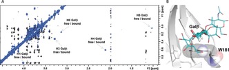
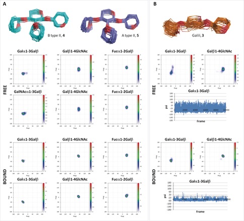
Ligand conformational entropy plays an important role in carbohydrate recognition events. Glycans are characterized by intrinsic flexibility around the glycosidic linkages, thus in most cases, loss of conformational entropy of the sugar upon complex formation strongly affects the entropy of the binding process. By employing a multidisciplinary approach combining structural, conformational, binding energy, and kinetic information, we investigated the role of conformational entropy in the recognition of the histo blood-group antigens A and B by human galectin-3, a lectin of biomedical interest. We show that these rigid natural antigens are pre-organized ligands for hGal-3, and that restriction of the conformational flexibility by the branched fucose (Fuc) residue modulates the thermodynamics and kinetics of the binding process. These results highlight the importance of glycan flexibility and provide inspiration for the design of high-affinity ligands as antagonists for lectins.
Keywords: blood-group antigen; conformational entropy; glycans; lectins; molecular recognition.
© 2019 Wiley-VCH Verlag GmbH & Co. KGaA, Weinheim.
Conflict of interest statement
The authors declare no conflict of interest.
Figures
Similar articles
-
Protein flexibility and conformational entropy in ligand design targeting the carbohydrate recognition domain of galectin-3.J Am Chem Soc. 2010 Oct 20;132(41):14577-89. doi: 10.1021/ja105852y. J Am Chem Soc. 2010. PMID: 20873837 Free PMC article.
-
Epitope mapping of histo blood group antigens bound to norovirus VLPs using STD NMR experiments reveals fine details of molecular recognition.Glycoconj J. 2017 Oct;34(5):679-689. doi: 10.1007/s10719-017-9792-5. Epub 2017 Aug 19. Glycoconj J. 2017. PMID: 28823097
-
Conformational dynamics and thermodynamics of protein-ligand binding studied by NMR relaxation.Biochem Soc Trans. 2012 Apr;40(2):419-23. doi: 10.1042/BST20110750. Biochem Soc Trans. 2012. PMID: 22435823 Review.
-
Interplay between Conformational Entropy and Solvation Entropy in Protein-Ligand Binding.J Am Chem Soc. 2019 Feb 6;141(5):2012-2026. doi: 10.1021/jacs.8b11099. Epub 2019 Jan 23. J Am Chem Soc. 2019. PMID: 30618244
-
Key regulators of galectin-glycan interactions.Proteomics. 2016 Dec;16(24):3111-3125. doi: 10.1002/pmic.201600116. Proteomics. 2016. PMID: 27582340 Free PMC article. Review.
Cited by
-
NMR Investigation of Protein-Carbohydrate Interactions: The Recognition of Glycans by Galectins Engineered with Fluorotryptophan Residues.Chemistry. 2023 Jan 24;29(5):e202202208. doi: 10.1002/chem.202202208. Epub 2022 Dec 12. Chemistry. 2023. PMID: 36343278 Free PMC article.
-
Oligosaccharide Presentation Modulates the Molecular Recognition of Glycolipids by Galectins on Membrane Surfaces.Pharmaceuticals (Basel). 2022 Jan 26;15(2):145. doi: 10.3390/ph15020145. Pharmaceuticals (Basel). 2022. PMID: 35215258 Free PMC article.
-
Regioselective Glycosylation Strategies for the Synthesis of Group Ia and Ib Streptococcus Related Glycans Enable Elucidating Unique Conformations of the Capsular Polysaccharides.Chemistry. 2019 Dec 18;25(71):16277-16287. doi: 10.1002/chem.201903527. Epub 2019 Nov 4. Chemistry. 2019. PMID: 31506992 Free PMC article.
-
Targeting Galectins With Glycomimetics.Front Chem. 2020 Aug 7;8:593. doi: 10.3389/fchem.2020.00593. eCollection 2020. Front Chem. 2020. PMID: 32850631 Free PMC article. Review.
-
Structure of a protective epitope reveals the importance of acetylation of Neisseria meningitidis serogroup A capsular polysaccharide.Proc Natl Acad Sci U S A. 2020 Nov 24;117(47):29795-29802. doi: 10.1073/pnas.2011385117. Epub 2020 Nov 6. Proc Natl Acad Sci U S A. 2020. PMID: 33158970 Free PMC article.
References
-
- Cummings R. D., Schnaar R. L., Esko J. D., Drickamer K., Taylor M. E., Essentials of Glycobiology, 3rd ed. (Eds.: A. Varki, R. D. Cummings, J. D. Esko, P. Stanley, G. W. Hart, M. Aebi, A. G. Darvill, T. Kinoshita, N. H. Packer, J. H. Prestegard, R. L. Schnaar, P. H. Seeberger), Cold Spring Harbor Laboratory Press, Cold Spring Harbor, 2015. –2017. - PubMed
-
- None
-
- Binder F. P. C., Lemme K., Preston R. C., Ernst B., Angew. Chem. Int. Ed. 2012, 51, 7327–7331; - PubMed
- Angew. Chem. 2012, 124, 7440–7444, and references therein;
-
- Topin J., Lelimousin M., Arnaud J., Audfray A., Pérez S., Varrot A., Imberty A., ACS Chem. Biol. 2016, 11, 2011–2020. - PubMed
Publication types
MeSH terms
Substances
Grants and funding
- CTQ2015-64597-C2-1P/Agencia Estatal de Investigación/International
- CTQ2015-64597-C2-2P/Agencia Estatal de Investigación/International
- CTQ2014-57141-R/Agencia Estatal de Investigación/International
- RYC-2014-15969/Ministerio de Economía, Industria y Competitividad, Gobierno de España/International
- Severo Ochoa EXcellence Accrediation/Ministerio de Economía, Industria y Competitividad, Gobierno de España/International
LinkOut - more resources
Full Text Sources
