. 2019 May;9(5):996-1007.
doi: 10.1002/2211-5463.12631. Epub 2019 Apr 3.

Incorporation of desmocollin-2 into the plasma membrane requires N-glycosylation at multiple sites

Affiliations

Incorporation of desmocollin-2 into the plasma membrane requires N-glycosylation at multiple sites

Andreas Brodehl et al. FEBS Open Bio. 2019 May.

Abstract

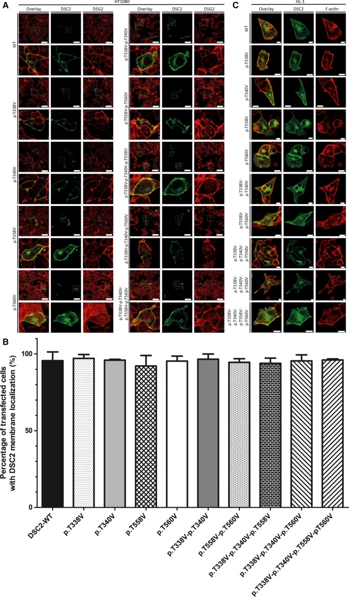
Desmocollin-2 (DSC2) is a desmosomal protein of the cadherin family. Desmosomes are multiprotein complexes, which are involved in cell adhesion of cardiomyocytes and of keratinocytes. The molecular structure of the complete extracellular domain (ECD) of DSC2 was recently described, revealing three disulfide bridges, four N-glycosylation sites, and four O-mannosylation sites. However, the functional relevance of these post-translational modifications for the protein trafficking of DSC2 to the plasma membrane is still unknown. Here, we generated a set of DSC2 mutants, in which we systematically exchanged all N-glycosylation sites, O-mannosylation sites, and disulfide bridges within the ECD and investigated the resulting subcellular localization by confocal laser scanning microscopy. Of note, all single and double N-glycosylation- deficient mutants were efficiently incorporated into the plasma membrane, indicating that the absence of these glycosylation sites has a minor effect on the protein trafficking of DSC2. However, the exchange of multiple N-glycosylation sites resulted in intracellular accumulation. Colocalization analysis using cell compartment trackers revealed that N-glycosylation- deficient DSC2 mutants were retained within the Golgi apparatus. In contrast, elimination of the four O-mannosylation sites or the disulfide bridges in the ECD has no obvious effect on the intracellular protein processing of DSC2. These experiments underscore the importance of N-glycosylation at multiple sites of DSC2 for efficient intracellular transport to the plasma membrane.

Keywords: N-glycosylation; O-mannosylation; arrhythmogenic (right ventricular) cardiomyopathy; desmocollin-2; desmosomes; vesicle transport.


Conflict of interest statement

The authors declare no conflict of interest.

Figures

Figure 1
Figure 1
Structural overview about the ECD of DSC2. (A) Molecular structures of the EC1 (5J5J) and EC2‐5 fragments of DSC2 (5ERP, https://www.rcsb.org) 10. The sugar residues of the N‐glycosylation sites are shown in dark blue, and the mannose residues of the O‐mannosylation sites are shown in light blue. The protein backbone is colored in green, Ca2+ ions are shown in orange, and cysteine residues forming disulfide bridges are labeled yellow. (B‐E) Schematic overviews about the generated DSC2 constructs. N‐glycosylation sites are shown in red, and O‐mannosylation sites are labeled in blue. Disulfide bridges are indicated by yellow and red lines. (B) DSC2 wild‐type construct. (C) N‐glycosylation‐ deficient DSC2 constructs. (D) O‐mannosylation‐ deficient constructs. (E) Disulfide bridge‐deficient DSC2 constructs.
Figure 2
Figure 2
Cellular localization of N‐glycosylation‐ deficient DSC2 mutants. (A) Representative fluorescence images and corresponding magnifications of transfected HT1080 cells expressing wild‐type DSC2‐eYFP or N‐glycosylation‐ deficient mutants are shown (green). Endogenous DSG2 was stained using anti‐DSG2 antibodies and is shown in red. Scale bars represent 50 μm. (B) Quantitative analysis of transfected cells with DSC2 membrane localization. Nonparametric Kruskal–Wallis test was used for statistical analysis. ***P < 0.001; n = 6. Error bars indicate mean ± SD. (C) Representative fluorescence images of transfected HL‐1 cells expressing wild‐type DSC2‐eYFP or N‐glycosylation‐ deficient mutants are shown (green). Endogenous F‐actin was stained using phalloidin conjugated to Texas Red. Scale bars represent 10 μm. White arrows indicate localization within the Golgi apparatus, and gray arrowheads indicate plasma membrane localization.
Figure 3
Figure 3
Cellular localization of O‐mannosylation‐ deficient DSC2 mutants. (A) Representative fluorescence images of transfected HT1080 cells expressing wild‐type DSC2‐eYFP or O‐mannosylation‐ deficient mutants are shown (green). Endogenous desmoglein‐2 is shown in red. Scale bars represent 50 μm. (B) Quantification of transfected HT1080 cells with DSC2 membrane localization revealed no significant differences between wild‐type and disulfide bridge‐deficient DSC2 mutants. Nonparametric Kruskal–Wallis test was used for statistical analysis. n = 3. Error bars indicate mean ± SD. (C) Representative fluorescence images of transfected HL‐1 cells expressing wild‐type DSC2‐eYFP (green) and disulfide bridge‐deficient mutants are shown. Endogenous F‐actin is labeled with phalloidin conjugated with Texas Red (red). Scale bars represent 10 μm.
Figure 4
Figure 4
Cellular localization of disulfide bridge‐deficient DSC2 mutants. (A) Representative fluorescence images of transfected HT1080 cells expressing wild‐type DSC2‐eYFP or disulfide bridge‐deficient mutants are shown (green). Endogenous desmoglein‐2 labeled with antibodies is shown in red. Scale bars represent 50 μm. (B) Quantification of transfected HT1080 cells with DSC2 membrane localization revealed no significant differences between wild‐type and disulfide bridge‐deficient DSC2 mutants. Nonparametric Kruskal–Wallis test was used for statistical analysis. n = 3. Error bars indicate mean ± SD. (C) Representative fluorescence images of transfected HL‐1 cells expressing wild‐type DSC2‐eYFP or disulfide bridge‐deficient mutants are shown (green). Endogenous F‐actin is labeled with phalloidin conjugated with Texas Red (red). Scale bars represent 10 μm.
Figure 5
Figure 5
Colocalization analysis of compartment trackers and the N‐glycosylation‐ deficient DSC2 mutants. HT1080 cells were transfected with (A) wild‐type and (B) mutant DSC2‐p.N166Q‐p.N392Q‐p.N546Q‐p.N629Q‐p.T338V‐p.T340‐p.T558V‐p.T560V constructs. Cell organelles were stained using compartment trackers. Colocalization of DSC2‐eYFP and the compartment trackers were evaluated using Leica Application Suite X software. Pearson's correlation of DSC2 mutant: 0.83; overlap coefficient: 0.88; colocalization rate: 85.4%. Scale bars represent 10 μm. (C) Schematic overview. ER, endoplasmic reticulum; G, Golgi apparatus; N, nucleus; PM, plasma membrane. (Images for ER and G are licenced from shutterstock.de)

References

    1. Patel DM and Green KJ (2014) Desmosomes in the heart: a review of clinical and mechanistic analyses. Cell Commun Adhes 21, 109–128. - PubMed
    1. Nomura T, Mizuno O, Miyauchi T, Suzuki S, Shinkuma S, Hata H, Fujita Y, Akiyama M and Shimizu H (2015) Striate palmoplantar keratoderma: report of a novel DSG1 mutation and atypical clinical manifestations. J Dermatol Sci 80, 223–225. - PubMed
    1. McKoy G, Protonotarios N, Crosby A, Tsatsopoulou A, Anastasakis A, Coonar A, Norman M, Baboonian C, Jeffery S and McKenna WJ (2000) Identification of a deletion in plakoglobin in arrhythmogenic right ventricular cardiomyopathy with palmoplantar keratoderma and woolly hair (Naxos disease). Lancet 355, 2119–2124. - PubMed
    1. Norgett EE, Hatsell SJ, Carvajal‐Huerta L, Cabezas JC, Common J, Purkis PE, Whittock N, Leigh IM, Stevens HP and Kelsell DP (2000) Recessive mutation in desmoplakin disrupts desmoplakin‐intermediate filament interactions and causes dilated cardiomyopathy, woolly hair and keratoderma. Hum Mol Genet 9, 2761–2766. - PubMed
    1. Klauke B, Kossmann S, Gaertner A, Brand K, Stork I, Brodehl A, Dieding M, Walhorn V, Anselmetti D, Gerdes D et al (2010) De novo desmin‐mutation N116S is associated with arrhythmogenic right ventricular cardiomyopathy. Hum Mol Genet 19, 4595–4607. - PubMed

Publication types