

Restoration of wall shear stress in the cephalic vein during extreme hemodynamics
- PMID: 30942634
- PMCID: PMC6714973
- DOI: 10.1080/03091902.2019.1591534
Restoration of wall shear stress in the cephalic vein during extreme hemodynamics
Abstract
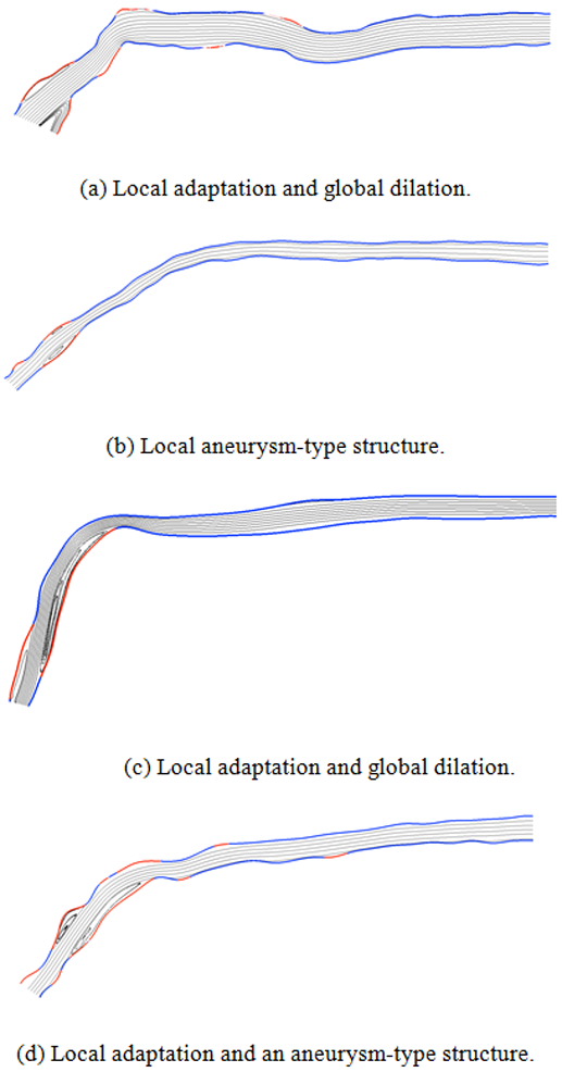
The surgical creation of an artery-vein connection via a Brachicephalic fistula (BCF) in patients with end stage renal disease (ESRD) provides a unique opportunity to study blood vessel response mechanisms to extreme hemodynamic conditions in relatively short timeframes. After BCF creation, the flow rate in the vein increases by an order of magnitude leading to separated flows and corresponding abnormally low, or negative, wall shear stress (WSS) in the curved arch segment of the cephalic vein. Locations of abnormally low WSS are shown to correlate with development of neointimal hyperplasia (NH) and subsequent stenosis. It is found that the stenosis, prior to a surgical intervention, restores the normal physiological WSS in the vein. As a result, this investigation provides evidence that the adaptation principle, known to apply in the arterial system, is also valid in the venous system. A novel graphical method is developed that combines clinical and computational data to assist in interpreting these physiological mechanisms including adaptation that lead to changes in vein geometry over time.
Keywords: Cephalic; brachiocephalic; fistula; vein adaptation; wall shear stress.
Conflict of interest statement
Disclosure statement
The authors report no conflicts of interest. No benefits in any form have been received from a commercial party related directly or indirectly to the subject of this manuscript.
Figures


Similar articles
-
A cohort study showing correspondence of low wall shear stress and cephalic arch stenosis in brachiocephalic arteriovenous fistula access.J Vasc Access. 2021 May;22(3):380-387. doi: 10.1177/1129729820942048. Epub 2020 Jul 21. J Vasc Access. 2021. PMID: 32693668
-
Hemodynamics in the cephalic arch of a brachiocephalic fistula.Med Eng Phys. 2014 Jul;36(7):822-30. doi: 10.1016/j.medengphy.2014.03.001. Epub 2014 Mar 30. Med Eng Phys. 2014. PMID: 24695337 Free PMC article.
-
Modified no-touch technique for radio-cephalic arteriovenous fistula increases primary patency and decreases juxta-anastomotic stenosis.J Vasc Access. 2024 May;25(3):904-913. doi: 10.1177/11297298221139339. Epub 2022 Dec 15. J Vasc Access. 2024. PMID: 36519744
-
A Review of the Hemodynamic Factors Believed to Contribute to Vascular Access Dysfunction.Cardiovasc Eng Technol. 2017 Sep;8(3):280-294. doi: 10.1007/s13239-017-0307-0. Epub 2017 May 19. Cardiovasc Eng Technol. 2017. PMID: 28527110 Review.
-
Role of Hypoxia and Metabolism in the Development of Neointimal Hyperplasia in Arteriovenous Fistulas.Int J Mol Sci. 2019 Oct 29;20(21):5387. doi: 10.3390/ijms20215387. Int J Mol Sci. 2019. PMID: 31671790 Free PMC article. Review.
Cited by
-
Computational modeling of the cephalic arch predicts hemodynamic profiles in patients with brachiocephalic fistula access receiving hemodialysis.PLoS One. 2021 Jul 14;16(7):e0254016. doi: 10.1371/journal.pone.0254016. eCollection 2021. PLoS One. 2021. PMID: 34260609 Free PMC article.
-
Creating patient-specific vein models to characterize wall shear stress in hemodialysis population.Comput Struct Biotechnol J. 2022 Oct 13;20:5729-5739. doi: 10.1016/j.csbj.2022.10.010. eCollection 2022. Comput Struct Biotechnol J. 2022. PMID: 36382195 Free PMC article.
-
Cephalic arch stenosis: an analysis of outcome by type of first intervention.CVIR Endovasc. 2024 Jan 19;7(1):13. doi: 10.1186/s42155-023-00424-4. CVIR Endovasc. 2024. PMID: 38240913 Free PMC article.
-
A novel red blood cell deformability biomarker is associated with hemolysis and vaso-occlusive crises in sickle cell disease.Sci Rep. 2025 May 7;15(1):15864. doi: 10.1038/s41598-025-00152-w. Sci Rep. 2025. PMID: 40335570 Free PMC article.
References
-
- Miller P, Tulwani A, Lusey C, and et al., 1999. “Predictors of adequacy of arteriovenous fistulas in hemodialysis patients”. Kidney Int, 56, pp. 275–286. - PubMed
-
- Rodriguez J, Armandans L, Ferrer E, Olmos A, Cordina S, Bartolome J, Borrelas J, and Piers L, 2000. “The function of permanent vascular access”. Nephrol Dial Transplant, 15, pp. 402–408. - PubMed
-
- Hammes M, Funaki B, and Coe F, 2008. “Cephalic arch stenosis in patients with fistula access for hemodialysis: relationship to diabetes and thrombosis”. Hemodial Int, 12, pp. 85–89. - PubMed
-
- Jaberi A, Schwartz D, Marticorena R, Dacouris N, Prabhudesai V, McFarlane P, and Donnelly S, 2007. “Risk factors for the development of cephalic arch stenosis”. J Vasc Access, 8, pp. 287–295. - PubMed
-
- Sivananthan G, Menashe L, and Halin N, 2014. “Cephalic arch stenosis in dialysis patients: review of clinical relevance, anatomy, current theories on etiology and management”. J Vasc Access, 15, pp. 157–162. - PubMed
Publication types
MeSH terms
Grants and funding
LinkOut - more resources
Full Text Sources
Medical
Research Materials