. 2019 May 9;62(9):4467-4482.
doi: 10.1021/acs.jmedchem.8b01986. Epub 2019 Apr 18.

Use of the 4-Hydroxytriazole Moiety as a Bioisosteric Tool in the Development of Ionotropic Glutamate Receptor Ligands

Affiliations

Use of the 4-Hydroxytriazole Moiety as a Bioisosteric Tool in the Development of Ionotropic Glutamate Receptor Ligands

Stefano Sainas et al. J Med Chem. .

Abstract

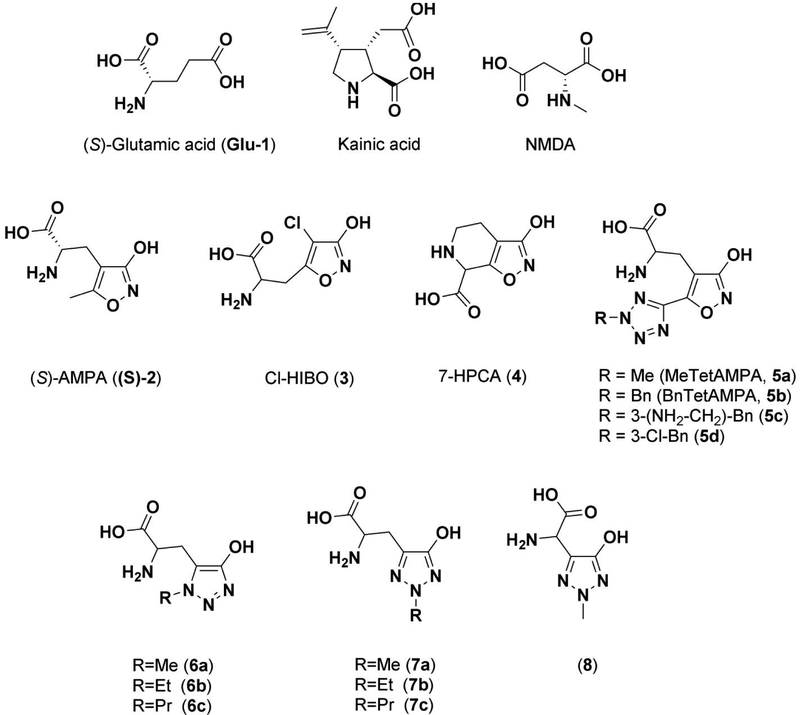
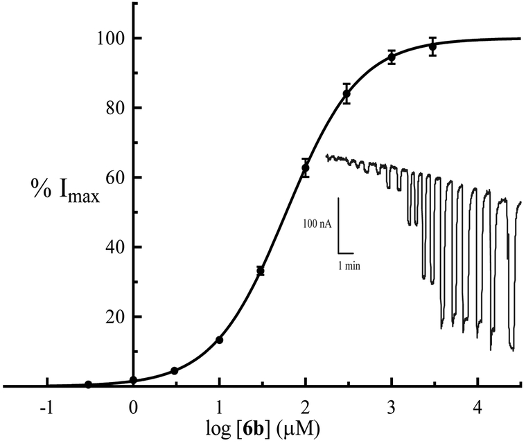
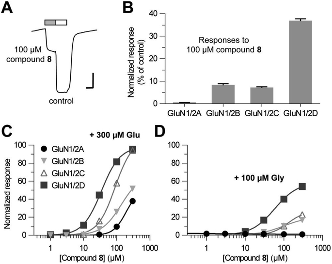
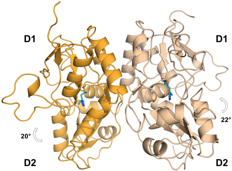
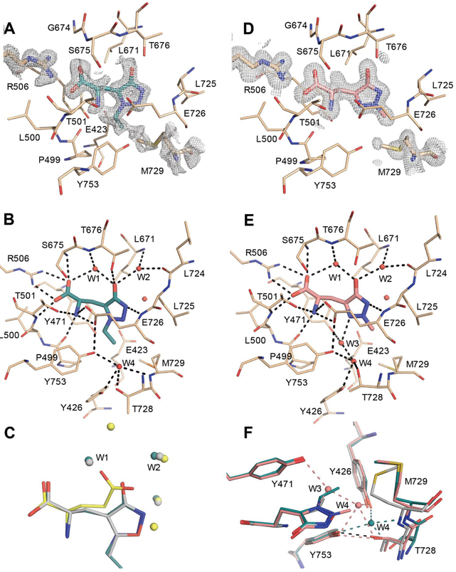
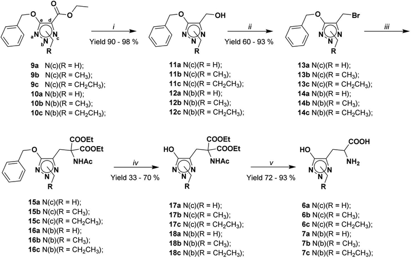
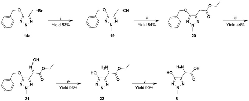
We report a series of glutamate and aspartate analogues designed using the hydroxy-1,2,3-triazole moiety as a bioisostere for the distal carboxylic acid. Compound 6b showed unprecedented selectivity among ( S)-2-amino-3-(3-hydroxy-5-methyl-4-isoxazolyl)propionic acid (AMPA) receptor subtypes, confirmed also by an unusual binding mode observed for the crystal structures in complex with the AMPA receptor GluA2 agonist-binding domain. Here, a methionine (Met729) was highly disordered compared to previous agonist-bound structures. This observation provides a possible explanation for the pharmacological profile. In the structure with 7a, an unusual organization of water molecules around the bioisostere arises compared to previous structures of ligands with other bioisosteres. Aspartate analogue 8 with the hydroxy-1,2,3-triazole moiety directly attached to glycine was unexpectedly able to activate both the glutamate and glycine agonist-binding sites of the N-methyl-d-aspartic acid receptor. These observations demonstrate novel features that arise when employing a hydroxytriazole moiety as a bioisostere for the distal carboxylic acid in glutamate receptor agonists.


Conflict of interest statement

CONFLICTS OF INTEREST

K.B.H. is a principal investigator on a research grant to University of Montana from Janssen Research & Development.

Figures

Figure 1.
Figure 1.
Structures of Glutamic acid, Kainic acid, NMDA, selective AMPA analogues and hydroxy-1,2,3-triazole analogues 6a-c, 7a-c and 8.
Figure 2.
Figure 2.
Normalized, averaged concentration-response data at homomeric rat GluA2(Q)i receptors expressed in Xenopus oocytes. Data are given as means ± SEM values of the pooled data. Responses from each oocyte were normalized to the maximum response of each oocyte before averaging. The top of the curve is fixed to 100% and the bottom to 0%. EC50 = 65 ± 6 μM, Hill slope = 1.07 ± 0.08 (n = 6 oocytes). Inset: Representative two-electrode voltage-clamp recording (Vh = −60 mV) with duplicate stimulations (0.3 – 1000 μM) followed by one stimulation at 3000 μM and one final stimulation of 1000 μM Glu.
Figure 3.
Figure 3.
(A) Representative two-electrode voltage-clamp recording of responses from recombinant GluN1/2D receptors expressed in Xenopus oocytes. Reponses were activated by 100 μM compound 8 as indicated by the grey bar, and control responses were activated by co-application of 300 μM Glu plus 100 μM Gly. The horizontal scale bars indicate 30 sec and the vertical scale bars indicate 200 nA. (B) Summary of responses to 100 μM compound 8 alone as percentage of control at recombinant GluN1/2A-D receptors. Data are given as mean ± SEM values from 4-6 oocytes. (C, D) Concentration-response data for compound 8 at recombinant NMDA receptor subtypes measured using two-electrode voltage-clamp recordings in the continuous presence of either 300 μM Glu (C) or 100 μM Gly (D). Responses are normalized to maximal activation by 300 μM Glu plus 100 μM Gly. Data are given as mean ± SEM values from 4 oocytes.
Figure 4.
Figure 4.
Structure of GluA2-ABD with 6b (PDB ID: 6Q54). The GluA2-ABD dimer is shown in cartoon representation with chain A in beige and chain B in orange. A grey arrow indicates D1-D2 domain closure. Molecule 6b is shown in cyan sticks representation.
Figure 5.
Figure 5.
Structures of GluA2-ABD with 6b (PDB ID: 6Q54) and 7a (PDB ID: 6Q60). (A) Simple PHENIX omit 2Fo–Fc map around 6b, Arg506 and Met729, carved at 1.6 Å and contoured at 1 sigma (chain A). GluA2-ABD residues within 4 Å of 6b are shown in beige sticks representation. Molecule 6b is shown in cyan sticks representation. (B) Hydrogen-bonding network (up to 3.2 Å; black dashed lines) among binding-site residues, 6b and water molecules (red spheres). (C) Comparison of binding mode of 6b (chain A), AMPA (grey; PDB code 1FTM, chain A) and Glu (yellow; PDB code 1FTJ, chain A). The structures were overlaid on lobe D1 residues. Only water molecules (within 4 Å of the ligands) are shown for clarity and colored according to the respective ligands. (D) Simple PHENIX omit 2Fo-Fc map around 7a, Arg506 and Met729, carved at 1.6 Å and contoured at 1 sigma (chain A). Molecule 7a is shown in salmon sticks representation. (E) Hydrogen bonding network among binding site residues, 7a and water molecules. (F) Zoom on the hydrogen bonding network of W3 and W4 in the GluA2-ABD structures with 6b (cyan), 7a (magenta) and AMPA (grey).
Scheme 1.
Scheme 1.
Synthesis of hydroxy-1,2,3-triazole Glu analogues 6a-c and 7a-c: i) NaBH4, EtOH abs; ii) NBS, (Ph)3P, dry DCM, −10°C; iii) diethyl-2-acetamidomalonate, NaH, dry THF; iv) H2, Pd/C 10% w/w, dry THF; v) a) 6N HCl, reflux; b) strong acidic cation exchange resin (elution with water until neutral pH. Finally the compound was eluted with 10% NH3 solution).
Scheme 2.
Scheme 2.
Synthesis of compound 8: i) NaCN, EtOH / H2O 9:1 v/v; ii) TMSCl, abs EtOH, 50 °C; iii) NaH, dry ethanol, EtONO; iv) a) H2, Pd/C, dry ethanol; b) HCl gas, dry ethanol; v) a) 6N HCl, reflux; b) strong acidic cation exchange resin (elution with water until neutral pH, then recover of the desired compound by elution with 10% w/w NH3 solution).

References

    1. Brauner-Osborne H; Egebjerg J; Nielsen EO; Madsen U; Krogsgaard-Larsen P Ligands for Glutamate Receptors: Design and Therapeutic Prospects. J Med Chem 2000, 43, 2609–2645. - PubMed
    1. Traynelis SF; Wollmuth LP; McBain CJ; Menniti FS; Vance KM; Ogden KK; Hansen KB; Yuan H; Myers SJ; Dingledine R Glutamate Receptor Ion Channels: Structure, Regulation, and Function. Pharmacol Rev 2010, 62, 405–496. - PMC - PubMed
    1. Paoletti P; Bellone C; Zhou Q Nmda Receptor Subunit Diversity: Impact on Receptor Properties, Synaptic Plasticity and Disease. Nat Rev Neurosci 2013, 14, 383–400. - PubMed
    1. Zhou Q; Sheng M Nmda Receptors in Nervous System Diseases. Neuropharmacology 2013, 74, 69–75. - PubMed
    1. Lau CG; Zukin RS Nmda Receptor Trafficking in Synaptic Plasticity and Neuropsychiatric Disorders. Nat Rev Neurosci 2007, 8, 413–426. - PubMed

Publication types