. 2019 Jun 1;30(12):1544-1554.
doi: 10.1091/mbc.E17-10-0591. Epub 2019 Apr 3.

Yb body assembly on the flamenco piRNA precursor transcripts reduces genic piRNA production

Affiliations

Yb body assembly on the flamenco piRNA precursor transcripts reduces genic piRNA production

Olesya A Sokolova et al. Mol Biol Cell. .

Abstract

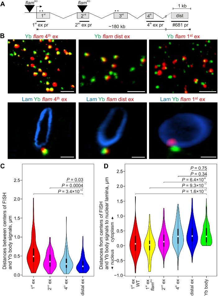
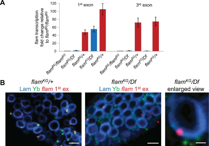
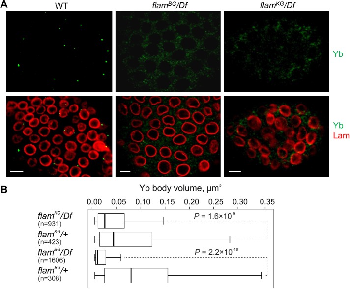
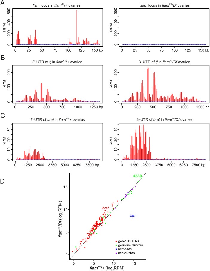
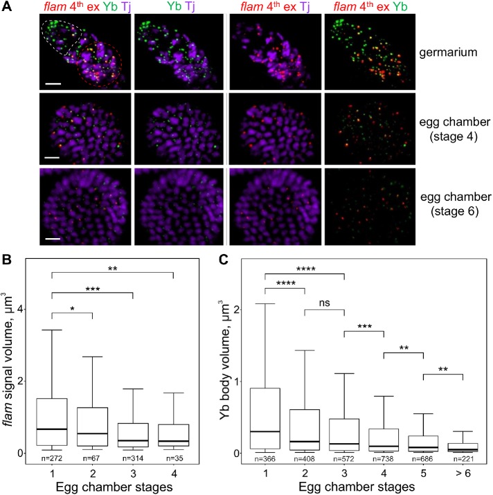
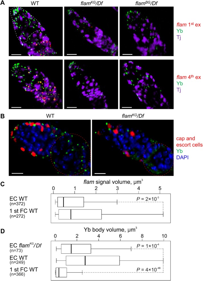
In Drosophila ovarian somatic cells, PIWI-interacting small RNAs (piRNAs) against transposable elements are mainly produced from the ∼180-kb flamenco (flam) locus. flam transcripts are gathered into foci, located close to the nuclear envelope, and processed into piRNAs in the cytoplasmic Yb bodies. The mechanism of Yb body formation remains unknown. Using RNA fluorescence in situ hybridization, we found that in the follicle cells of ovaries the 5'-ends of flam transcripts are usually located in close proximity to the nuclear envelope and outside of Yb bodies, whereas their extended downstream regions mostly overlap with Yb bodies. In flamKG mutant ovaries, flam transcripts containing the first and, partially, second exons but lacking downstream regions are gathered into foci at the nuclear envelope, but Yb bodies are not assembled. Strikingly, piRNAs from the protein-coding gene transcripts accumulate at higher levels in flamKG ovaries indicating that piRNA biogenesis may occur without Yb bodies. We propose that normally in follicle cells, flam downstream transcript regions function not only as a substrate for generation of piRNAs but also as a scaffold for Yb body assembly, which competitively decreases piRNA production from the protein-coding gene transcripts. By contrast, in ovarian somatic cap and escort cells Yb body assembly does not require flam transcription.


Figures

FIGURE 1:
FIGURE 1:
The fourth and distal exons of flam transcripts are mostly localized in Yb bodies, whereas the flam first exon is not. (A) Scheme of the flam locus with the designation of exon/intron structure (according to Goriaux et al. [2014] and FlyBase r6), sites of TE insertions in flamBG and flamKG mutations (black triangles), RNA FISH probes (black rectangles), and primer pairs used for RT-qPCR (arrowheads). (B) RNA FISH (red) with flam fourth exon probe (left panels), distal exon probe (middle panels), or first exon probe (right panels) coupled with immunostaining of Yb bodies with anti-Yb antibody (green; top panels) and nuclear envelope with anti-lamin Dm0 antibody (blue; enlarged view of representative nuclei; bottom panels) in the follicle cells of stage 12 egg chambers. Full Z-series projections of confocal image stacks are shown in the top panels, and single optical sections are shown in the bottom panels. Scale bars are 5 μm in the top panels and 1.5 μm in the bottom panels. (C) Violin plots showing the distribution of distances between centers of Yb bodies and flam RNA FISH signals for the first (red), second (magenta), fourth (blue), or distal (dark blue) exon probes in the follicle cells of early egg chambers. Distances were measured with IMARIS software for 40–50 cells in two biological replicates for each probe. (D) Violin plots showing the distribution of distances from the centers of flam RNA FISH signals and Yb bodies to the nuclear lamina. Distances for 40–50 cells in two biological replicates (for first, second, fourth, and distal exons in WT ovaries), in one replicate (for first exon in flamKG/Df ovaries), or in eight replicates (for Yb bodies) were measured with IMARIS software. In C and D M-W U-test was used for the pairwise comparison of distributions between the distances of the first exon-Yb, the second exon-Yb, and the fourth exon-Yb (C), or between the distances of the first, second, fourth, and distal exons and Yb bodies to the nuclear lamina (D).
FIGURE 2:
FIGURE 2:
flam first exon transcripts are retained and form perinuclear foci in flamKG/Df ovaries. (A) RT-qPCR analysis of flam transcription in flamBG and flamKG mutant ovaries using primer pairs for the first or third exons. Fold change of flam transcription in the ovaries with indicated genotypes relative to flamBG/flamBG ovaries, normalized on the Adh transcript levels, is indicated along the Y-axis (mean ± SD for two replicates). The flam third exon transcripts were barely detected in flamBG and flamKG mutants, but the flam first exon transcripts are retained in flamKG mutant. (B) RNA FISH (red) with flam first exon probe coupled with immunostaining of Yb bodies with anti-Yb antibody (green) and nuclear envelope with anti-lamin Dm0 antibody (blue) in the follicle cells of the stage 2 egg chamber of flamKG/+ (left panel), flamKG/Df (central panel), and enlarged view of flamKG/Df (right panel). Scale bars are 5 μm in the left and central panels and 0.7 μm in the right panel.
FIGURE 3:
FIGURE 3:
Disintegration of Yb bodies in flam mutants. (A) Immunostaining of follicle cells from fourth stage egg chambers in flamBG/+ (WT), flamBG/flamBG, and flamKG/Df ovaries with anti-Yb (green) and anti-lamin Dm0 (red) antibodies. Scale bars are 7 μm. (B) Box plots showing a distribution of volumes of Yb bodies from egg chambers of stages 3–5 measured with IMARIS software in flam mutants in comparison with the corresponding controls. The number of counted signals is indicated for each genotype. M-W U-test was used for the pairwise comparison of distributions between mutant and control ovaries.
FIGURE 4:
FIGURE 4:
piRNA production from the protein-coding gene transcripts is enhanced in flamKG ovaries. (A–C) piRNA density (in RPM) for uniquely mapped reads along the entire flam (A), 3′-UTR of tj (B), and 3′-UTR of brat (C) piRNA clusters. Sense and antisense piRNAs are indicated in red and blue, respectively. (D) Scatter plot showing an abundance of piRNAs (in log2 RPM) for somatic genic piRNA clusters (red dots), predominantly germline piRNA clusters (green dots), and for the top 10 microRNAs most abundant in OSCs (purple dots) in flamKG/+ (control) or flamKG/Df small RNA-seq libraries. The median fold change for somatic genic piRNA clusters and for predominantly germline piRNA clusters upon flamKG mutation is 1.6 and 1.1, respectively. The difference in piRNA production between “germline” and genic clusters is highly significant (P < 0.0001, M-W U-test).
FIGURE 5:
FIGURE 5:
In ovarian follicle cells flam foci as well as the Yb bodies become progressively smaller during egg chamber maturation. (A) RNA FISH with flam fourth exon probe (red) in WT (flamKG/+) ovaries coupled with the immunostaining of Yb bodies by anti-Yb antibody (green) and anti-Tj antibody (violet), the nuclear marker of OSCs. Egg chambers at different stages in the same ovariole are shown. The apical region of a germarium is populated by cap and escort cells (outlined by a white dotted line), and the first stage follicle cells are located distally (outlined by a red dotted line). flam foci are most prominent in the first stage follicle cells of germaria (top panel) as well as in stage 2–3 egg chambers, whereas at the later stages of oogenesis (middle and bottom panels) flam as well as Yb body signals become weaker. Scale bars are 10 μm in the top panel and 7 μm in the middle and bottom panels. (B, C) Box plots showing a distribution of volumes of the flam fourth exon (B) or the Yb body (C) signals at different stages of egg chamber maturation in WT (flamKG/+) ovaries. Volumes of flam foci or Yb bodies from the indicated stages were measured with IMARIS software. The number of counted signals is indicated for each stage. M-W U-test was used for the pairwise comparison of distributions. *, P < 0.05; **, P < 0.01; ***, P < 0.001; ****, P < 0.0001; ns, not significant.
FIGURE 6:
FIGURE 6:
In cap and escort cells the assembly of Yb bodies does not depend on flam transcription. (A) RNA FISH with flam first (top panel) or fourth (bottom panel) exon probes (red) coupled with Yb (green) and Tj (violet) immunostaining. In cap and escort cells (outlined by a white dotted line in germarium) of WT (flamKG/+) ovaries (left panel) flam RNA FISH signals are weaker than in the distally located first egg chamber follicle cells, while the Yb bodies are more strongly stained. In cap and escort cells of flam mutants (middle and right panels) Yb bodies are assembled. Scale bars are 10 μm. (B) Germaria of PZ1444 (WT, left panel) or flamKG/Df; PZ1444 (right panel) ovaries (PZ1444 is a lacZ enhancer trap line that is expressed in the cap and escort cells; Margolis and Spradling, 1995; Xie and Spradling, 2000) immunostained with anti-Yb (green) and anti–β-galactosidase (red) antibodies, counterstained with DAPI (blue). Yb bodies are larger in the cap and escort cells (their nuclei are marked by red) than in the follicle cells (outlined by a red dotted line). In flamKG/Df ovaries Yb bodies are disintegrated in follicle cells, but not in the cap and escort cells. Scale bars are 10 μm. (C, D) Box plots showing a distribution of volumes of flam fourth exon (C) or Yb body (D) signals in the cap and escort cells (EC) or in the first stage egg chamber follicle cells (1 st FC) of WT (flamKG/+) or flamKG/Df ovaries. The number of counted signals is indicated for each cell type. M-W U-test was used for the pairwise comparison of distributions.

References

    1. Brennecke J, Aravin AA, Stark A, Dus M, Kellis M, Sachidanandam R, Hannon GJ. (2007). Discrete small RNA-generating loci as master regulators of transposon activity in Drosophila . Cell , 1089–1103. - PubMed
    1. Chujo T, Hirose T. (2017). Nuclear bodies built on architectural long noncoding RNAs: unifying principles of their construction and function. Mol Cells , 889–896. - PMC - PubMed
    1. Czech B, Hannon GJ. (2016). One loop to rule them all: the ping-pong cycle and piRNA-guided silencing. Trends Biochem Sci , 324–337. - PMC - PubMed
    1. Dennis C, Brasset E, Sarkar A, Vaury C. (2016). Export of piRNA precursors by EJC triggers assembly of cytoplasmic Yb-body in Drosophila . Nat Commun , 13739. - PMC - PubMed
    1. Dennis C, Zanni V, Brasset E, Eymery A, Zhang L, Mteirek R, Jensen S, Rong YS, Vaury C. (2013). “Dot COM,” a nuclear transit center for the primary piRNA pathway in Drosophila . PLoS One , e72752. - PMC - PubMed

Publication types