. 2019 Apr 3;15(4):e1007680.
doi: 10.1371/journal.ppat.1007680. eCollection 2019 Apr.

USP49 negatively regulates cellular antiviral responses via deconjugating K63-linked ubiquitination of MITA

Affiliations

USP49 negatively regulates cellular antiviral responses via deconjugating K63-linked ubiquitination of MITA

Liya Ye et al. PLoS Pathog. .

Abstract

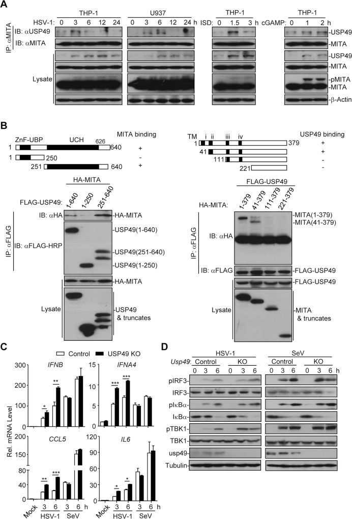
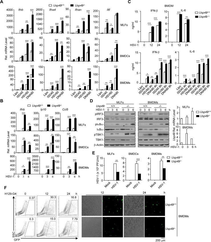
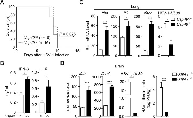
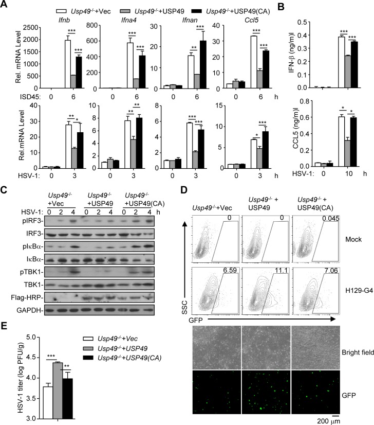
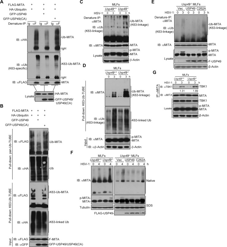
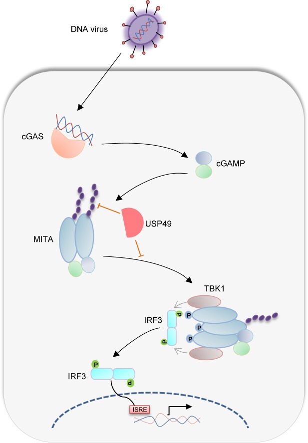
Mediator of IRF3 activation (MITA, also known as STING and ERIS) is an essential adaptor protein for cytoplasmic DNA-triggered signaling and involved in innate immune responses, autoimmunity and tumorigenesis. The activity of MITA is critically regulated by ubiquitination and deubiquitination. Here, we report that USP49 interacts with and deubiquitinates MITA after HSV-1 infection, thereby turning down cellular antiviral responses. Knockdown or knockout of USP49 potentiated HSV-1-, cytoplasmic DNA- or cGAMP-induced production of type I interferons (IFNs) and proinflammatory cytokines and impairs HSV-1 replication. Consistently, Usp49-/- mice exhibit resistance to lethal HSV-1 infection and attenuated HSV-1 replication compared to Usp49+/+ mice. Mechanistically, USP49 removes K63-linked ubiquitin chains from MITA after HSV-1 infection which inhibits the aggregation of MITA and the subsequent recruitment of TBK1 to the signaling complex. These findings suggest a critical role of USP49 in terminating innate antiviral responses and provide insights into the complex regulatory mechanisms of MITA activation.


Conflict of interest statement

The authors have declared that no competing interests exist.

Figures

Fig 1
Fig 1. USP49 interacts with MITA and inhibits MITA-mediated signaling.
(A) Immunoprecipitation (with anti-MITA) and immunoblot analysis (with anti-MITA or anti-USP49) of THP-1 or U937 cells infected with HSV-1, or transfected with ISD (10 μg) or cGAMP (4 μg) for the indicated time points. (B) Immunoprecipitation (with anti-Flag) and immunoblot analysis (with anti-FLAG or anti-HA) of HEK293 cells transfected with plasmids encoding HA-MITA and FLAG-tagged USP49 or USP49 truncates for 24 h or transfected with plasmids encoding FLAG-USP49 and HA-MITA or MITA truncates for 24 h. (C) qRT-PCR analysis of IFNB, IFNA4, CCL5 and IL6 in control and USP49 KO THP-1 cells infected with HSV-1 or SeV for 0–6 h. (D) Immunoblot analysis of phosphorylated and total IRF3, IκBα, TBK1 and Tubulin in control and USP49 KO THP-1 cells infected with HSV-1 or SeV for 0–6 h. *P < 0.05; **P < 0.01; ***P <0.001 (analysis of two-way ANOVA followed by Bonferroni post-test). Data are representative of three independent experiments (mean ± S.D. in C).
Fig 2
Fig 2. Knockout of USP49 potentiates HSV-1-triggered signaling.
(A) qRT-PCR analysis of Ifnb, Ifna4, lfnan and Il6 mRNA in Usp49+/+ and Usp49-/- MLFs (upper), BMDCs (middle) and BMDMs (lower) left mock transfected (Lipo) or transfected with ISD45, HSV60, DNA90 for 0–6 h. (B) qRT-PCR analysis of Ifnb, IP10,and Ccl5 mRNA in Usp49+/+ and Usp49-/- MLFs (upper), BMDCs (middle) and BMDMs (lower) infected with HSV-1 for 0–6 h. (C) ELISA analysis of IFN-β and IL-6 in the supernatants of Usp49+/+ and Usp49-/- BMDMs infected for 0–24 hours or mock transfected (Lipo) or transfected with ISD45, HSV60, DNA90 for 4–8 h. (D) Immunoblot analysis of phosphorylation of IRF3, IκBα, TBK1, total IRF3, IκBα, TBK1 and β-Actin in Usp49+/+ and Usp49-/- MLFs (left) and BMDCs (middle) infected with HSV-1 for 0–6 hours. qRT-PCR analysis of Usp49 mRNA in Usp49+/+ and Usp49-/- MLFs and BMDCs infected with HSV-1 for 0–6 h (right). (E) Plaque assay of HSV-1 replication in the supernatants of Usp49+/+ and Usp49-/- MLFs, BMDCs and BMDMs infected with HSV-1(MOI of 0.4) for 1 h followed by twice PBS wash and cultured in full medium for 60 h. (F) Flow cytometry analysis (left), microscopy imaging (right) of Usp49+/+ and Usp49-/- BMDMs infected with H129-G4 for 0–24 h. *P < 0.05; **P < 0.01; ***P <0.001 (analysis of two-way ANOVA followed by Bonferroni post-test). Data are representative of three independent experiments (mean ± S.D. in A-E).
Fig 3
Fig 3. Usp49-/- mice exhibit resistance to lethal HSV-1 infection.
(A) Survival (Kaplan-Meier curve) of Usp49+/+ (n = 16) and Usp49-/- (n = 16) intravenously injected with HSV-1 (2.5×10⁶ PFU per mouse) monitored survival for 14 days. (B) ELISA analysis of IFN-β and IL-6 in the serum of Usp49+/+ and Usp49-/- mice(n = 3) intravenously injected with HSV-1 (2.5×10⁶ PFU per mouse) for 12 hours. (C) qRT-PCR analysis of Ifnb, Il6, Ifnan and HSV-1-UL30 mRNA in the lungs (n = 3) from Usp49+/+ and Usp49-/- mice intravenously injected with HSV-1 (2.5×10⁶ PFU per mouse) for 24 hours. (D) qRT-PCR analysis of Ifnb, Ifna4, HSV-1-UL30 mRNA and plaque assay of the brains (n = 3) from Usp49+/+ and Usp49-/- mice intranasally injected with HSV-1 (2.5×10⁶ PFU per mouse) for 4 days. *P < 0.05; **P < 0.01; ***P <0.001 (two-tailed t-test). Data are representative of three independent experiments (mean ± S.D. in B-D).
Fig 4
Fig 4. The DUB activity of USP49 is required for inhibition of HSV-1-triggered signaling.
(A, B) qRT-PCR analysis of Ifnb, Ifna4, Ifnan and Ccl5 mRNA (A), and ELISA analysis of IFN-β, Ccl5 (B) in Usp49-/- mice MLFs reconstituted with empty vector (Usp49-/- +vec), Usp49 (Usp49-/- + Usp49) or Usp49CA (Usp49-/- + Usp49(CA)) infected with ISD45 for 0–6 hours (A), HSV-1 for 0–3 hours (A) or 0–10 hours (B). (C, D) Immunoblot (C), flow cytometry(D, upper graphs), microscopy imaging (D, lower graphs) and plaque assay (E) of Usp49-/- mice MLFs reconstituted with empty vector (Usp49-/- +vec), Usp49 (Usp49-/- + Usp49) or Usp49CA (Usp49-/- + Usp49(CA)) infected with HSV-1 for 0–4 hours (C), 12 hours (D) or 1 hour (E). *P < 0.05; **P < 0.01; ***P <0.001 (analysis of two-way ANOVA followed by Bonferroni post-test). Data are representative of two (C) or three (A, B, D, E) independent experiments (mean ± S.D. in A, B, E).
Fig 5
Fig 5. USP49 deubiquitinates and inhibits aggregation of MITA.
(A) Denature-immunoprecipitation (Denature-IP) (with anti-FLAG or IgG as a control) and immunoblot analysis (with anti-FLAG, anti-HA, anti-K63-linked ubiquitin or anti-GFP) of HEK293 cells transfected with plasmids encoding FLAG-MITA, HA-Ubiquitin and empty vector or GFP-USP49 or GFP-USP49 (CA) for 24 h. (B) Pulldown (with GST beads and GST-TUBEs) and immunoblot analysis (with anti-FLAG, anti-HA or anti-GFP) of HEK293 cells transfected with plasmids encoding FLAG-MITA, HA-Ubiquitin and empty vector or GFP-USP49 or GFP-USP49 (CA) for 24 h. (C) Denature-IP (with anti-MITA) and immunoblot analysis (anti-K63-linked ubiquitin, anti-MITA or anti-β-Actin) of Usp49+/+ and Usp49-/- MLFs infected with HSV-1 for 0–3 h. (D) Pulldown (with GST beads and TUBE) and immunoblot analysis (anti-K63-linked ubiquitin, anti-MITA or anti-β-Actin) of Usp49+/+ and Usp49-/- MLFs infected with HSV-1 for 0–3 h. (E) Denature-IP (with anti-MITA) and immunoblot analysis (with anti-K63-linked Ub, anti-MITA, anti-FLAG or anti- anti-β-Actin) of Usp49-/- MLFs reconstituted with empty vector, USP49 or USP49 (CA) infected with HSV-1 for 0–3 h. (F) Native–PAGE analysis and SDS–PAGE of the aggregation of MITA in Usp49+/+ and Usp49-/- MLFs or Usp49-/- MLFs reconstituted with empty vector, USP49 or USP49 (CA) infected with HSV-1 for 0–4 h. (G) Immunoprecipitation (with anti-MITA) and immunoblot analysis (with anti-TBK1, anti-MITA, anti-USP49 or anti-β-Actin) MLFs infected with HSV-1 for 0–4 h. Data are representative of at least three independent experiments.
Fig 6
Fig 6. A model on USP49-mediated regulation of antiviral responses.
HSV-1 infection induces production of cGAMP that binds to MITA and leads to K63-linked ubiquitination and oligomerization of MITA. USP49 removes K63-linked ubiquitin chains from MITA and thereby inhibits the oligomerization. Such a deubiquitination impairs the subsequent recruitment of TBK1 and phosphorylation of IRF3 after HSV-1 infection.

References

    1. Akira S, Uematsu S, Takeuchi O. Pathogen recognition and innate immunity. Cell. 2006;124(4):783–801. 10.1016/j.cell.2006.02.015 WOS:000237240900021. - DOI - PubMed
    1. Wu J, Chen ZJ. Innate Immune Sensing and Signaling of Cytosolic Nucleic Acids. In: Littman DR, Yokoyama WM, editors. Annual Review of Immunology, Vol 32 Annual Review of Immunology. 322014. p. 461–88. 10.1146/annurev-immunol-032713-120156 - DOI - PubMed
    1. Hu M-M, Shu H-B. Cytoplasmic Mechanisms of Recognition and Defense of Microbial Nucleic Acids. Annual review of cell and developmental biology. 2018. 10.1146/annurev-cellbio-100617-062903 MEDLINE:30095291. - DOI - PubMed
    1. Tan X, Sun L, Chen J, Chen ZJ. Detection of Microbial Infections Through Innate Immune Sensing of Nucleic Acids. Annual review of microbiology. 2018;72:447–78. 10.1146/annurev-micro-102215-095605 MEDLINE:30200854. - DOI - PubMed
    1. Ma Z, Ni G, Damania B. Innate Sensing of DNA Virus Genomes. Annual review of virology. 2018;5(1):341–62. 10.1146/annurev-virology-092917-043244 MEDLINE:30265633. - DOI - PMC - PubMed

Publication types

MeSH terms