Review
. 2019 Apr 2;29(4):803-826.
doi: 10.1016/j.cmet.2019.03.003.

Watch What You (Self-) Eat: Autophagic Mechanisms that Modulate Metabolism

Affiliations
Review

Watch What You (Self-) Eat: Autophagic Mechanisms that Modulate Metabolism

Vikramjit Lahiri et al. Cell Metab. .

Abstract

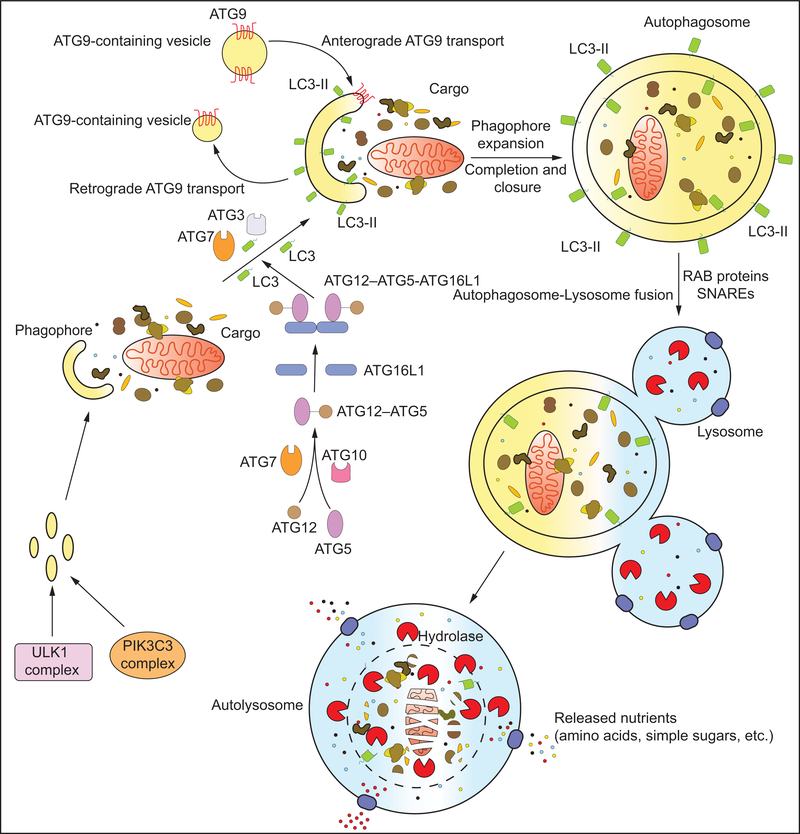
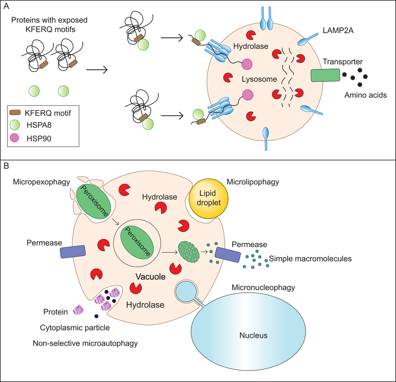
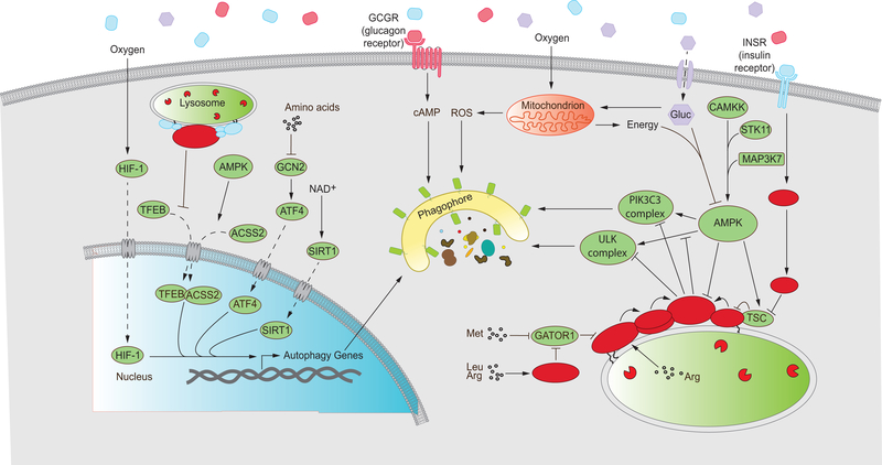
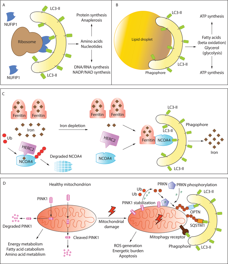
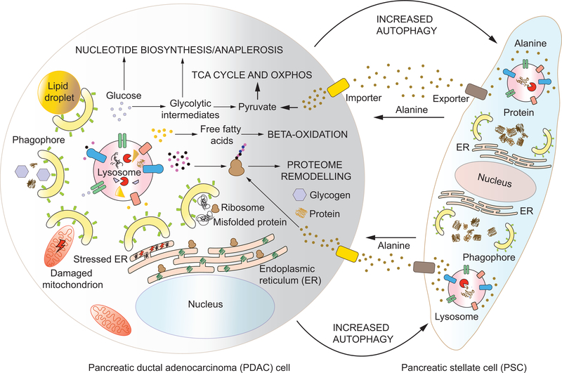
Autophagy is an evolutionarily conserved lysosome- or vacuole-dependent catabolic pathway in eukaryotes. Autophagy functions basally for cellular quality control and is induced to act as an alternative source of basic metabolites during nutrient deprivation. These functions of autophagy are intimately connected to the regulation of metabolism, and the metabolic status of the cell in turn controls the nature and extent of autophagic induction. Here, we highlight the co-regulation of autophagy and metabolism with a special focus on selective autophagy that, along with bulk autophagy, plays a central role in regulating and rewiring metabolic circuits. We outline the metabolic signals that activate these pathways, the mechanisms involved, and the downstream effects and implications while recognizing yet unanswered questions. We also discuss the role of autophagy in the development and maintenance of adipose tissue, an emerging player in systemic metabolic homeostasis, and describe what is currently known about the complex relationship between autophagy and cancer.

Keywords: AMPK; autophagy; ferritinophagy; homeostasis; lysosome; mTOR; macroautophagy; mitophagy; physiology; protein turnover; proteostasis; stress.


Conflict of interest statement

Declaration of Interest

The authors declare no competing interests.

Figures

Figure 1:
Figure 1:. The molecular machinery of macroautophagy:
The hallmark of macroautophagy is the double membrane autophagosome that forms by the de novo assembly of membrane from various sources. The process begins with the formation of the phagophore, a process initiated by the ULK1 and VPS34 complexes. Expansion of the phagophore occurs via the continued recruitment of membrane vesicles by ATG9 as well as the conjugation of LC3 to the phagophore membrane (to form LC3-II). LC3 conjugation involves a two-step ubiquitin-like conjugation pathway involving ATG7, ATG10, ATG3, ATG12, ATG5 and ATG16 (refer to text for details). The phagophore expands around the cargo, finally closing to form a cargo-containing autophagosome. The autophagosome subsequently fuses with lysosome(s) by the concerted action of Rab and SNARE proteins to form the autolysosome. Lysosomal hydrolases degrade the inner autophagosomal membrane and the enclosed cargo. The breakdown products, simple macromolecules such as amino acids, are subsequently transported out in the cytoplasm by lysosomal transporters for reuse.
Figure 2:
Figure 2:. Other mechanisms of self-eating: Chaperone-mediated autophagy (CMA) and microautophagy:
(A) In yeast, microautophagy involves the sequestration of cargo by the protrusion/invagination of the vacuolar membrane followed by an inward scission leading to the formation of a cargo-containing lumenal vesicle. This vesicle is subsequently degraded by vacuolar hydrolases releasing simple breakdown products. Microautophagy can be non-selective, degrading cytosolic components randomly, or selective, specifically degrading lipid droplets (microlipophagy) or peroxisomes (macropexophagy). Another selective microautophagic process, not discussed in the text, is the piecemeal microautophagy of the nucleus (PMN) which degrades portions of the nucleus. (B) CMA is a lysosome-dependent protein degradation pathway that requires the cytosolic chaperone HSC70. Proteins with an exposed KFERQ or KFERQ-like motif are recognized and bound by HSC70. The complex then locates to the lysosomal membrane where the multimerization of LAMP2A allows the formation of a conduit for the delivery of the protein into the lysosomal lumen, a process facilitated by the lumenal chaperone HSP90. Lysosomal hydrolases break down the protein releasing amino acids which are transported into the cytosol.
Figure 3:
Figure 3:. An intricate network of regulatory components and signaling pathways influence autophagy in response to cellular metabolic status.
Autophagy is regulated at multiple levels by cellular components that respond to specific or general metabolic cues. Several proteins such as ATF4, HIF-1, SIRT1 and TFEB modulate the expression of autophagy-related genes at the transcriptional level. These pathways are sensitive to the abundance of amino acids, oxygen availability, the reduction status of the cellular NAD pool and activation status of MTORC1 and AMPK. Expression of autophagy genes leads to autophagy induction, depicted as an expanding phagophore. Glucagon signals a fasted organismal status and upregulates autophagy through cAMP-dependent pathways. Glucose fuels oxidative phosphorylation in mitochondria, providing energy in the form of ATP but also generating ROS that indirectly upregulate autophagy. Low cellular energy charge activates AMPK in a process that requires upstream kinases such as CAMKK and STK11. AMPK promotes autophagy by activating the autophagy-initiating ULK1 and VPS34 complexes as well as inhibiting MTORC1 function. MTORC1 inhibits autophagy when recruited to the lysosome and activated. MTORC1 recruitment and activation occurs in response to the presence of both growth factors such as INS/insulin and an abundance of amino acids in the cytosol and lysosomal lumen. While INS signaling occurs through the PI3K-AKT-TSC axis, leading to the activation of the small GTPase RHEB, amino acid sufficiency is conveyed through the Ragulator complex that impinges on the small GTPases known as RRAGs. The RRAG complex represents a heterodimer between RRAGA (or B) and RRAGC (or D). Activated MTORC1 inhibits the ULK1 and VPS34 complexes to downregulate autophagy (Refer to text for details). Solid arrows represent upregulation, blunt arrows represent repression and dashed arrows represent movement/transport. Gluc, glucose; Met, methionine; Arg, Arginine; Leu, leucine.
Figure 4:
Figure 4:. Selective autophagy as a modulator of metabolic homeostasis:
Selective autophagy removes dysfunctional/superfluous organelles downstream of metabolic cues. It also provides a source of raw material for several metabolic processes and pathways. Selective autophagy involves the sequestration of specific cargo by a LIR-containing receptor that links the cargo with LC3-II (see text for details). An example in (A) shows the selective targeting of ribosomes to the mitochondria by the ribophagy receptor NUFIP1. Designated receptors have not yet been identified for all types of selective autophagy. Selective uptake of lipid droplets (B) may simply occur by the formation and expansion of the phagophore on the surface of the droplet. (C) Ferritinophagy allows the iron-dependent regulation of Ferritin degradation. NCOA4 is the receptor that targets the iron-storing protein Ferritin to LC3-II. Under conditions of ironsufficiency, NCOA4 is ubiquitinated by HERC2 via an iron-dependent interaction, leading to NCOA4 degradation. When the cellular levels of free iron decline, this interaction is weakened allowing NCOA4 to target Ferritin to the phagophore. The degradation of Ferritin releases free iron. (D) Healthy mitochondria are the principal source of cellular ATP and regulate multiple metabolic circuits. Damaged mitochondria, that are detrimental, are removed by mitophagy. In the PINK1-PRKN dependent pathway of mitophagy, the kinase PINK1 which is imported and cleaved in healthy mitochondria, and subsequently targeted for cytosolic degradation, is stabilized on the OMM (outer mitochondrial membrane). PINK1 phosphorylates ubiquitin and the E3 ubiquitin ligase PRKN promoting large-scale ubiquitination of mitochondrial OMM proteins. Ubiquitinated proteins are recognized by ubiquitin-binding autophagy adaptors such as OPTN and SQSTM1 which also bind LC3-II, promoting mitochondrial degradation (refer to text for details). Mitophagy may also be orchestrated by OMM/IMM (inner mitochondrial membrane) proteins that directly bind LC3-II and function as mitophagy receptors (refer to text and Table 1 for details).
Figure 5:
Figure 5:. Autophagy in tumor cells and the stroma sustains tumor progression:
Autophagy is a pro-tumorigenic pathway in transformed cells, helping them survive. Within cancer cells, autophagy removes detrimentally damaged mitochondria and helps relieve ER-stress. Autophagy is also responsible for the removal of toxic, misfolded proteins. The recycling of proteins, lipid droplets, glycogen and ribosomes by autophagy promotes energy metabolism and anabolic synthesis by providing substrates for metabolic pathways. In addition, certain cancer cells like pancreatic ductal adenocarcinoma (PDAC) cells induce autophagy in neighboring stromal cells, pancreatic stellate cells (PSCs) in the case of PDAC. The degradation of proteins by autophagy in PSCs promotes alanine production and secretion. PDACs import alanine, which may be channeled into protein synthesis or, more importantly, be converted to pyruvate. This allows an external source for pyruvate and subsequent mitochondrial energy production, thereby allowing PDACs to utilize glycolytic intermediates for nucleotide synthesis and anaplerotic reactions that fuel growth (see text for details).

References

    1. Adamska A, Domenichini A, and Falasca M (2017). Pancreatic Ductal Adenocarcinoma: Current and Evolving Therapies. Int J Mol Sci 18. - PMC - PubMed
    1. Altshuler-Keylin S, Shinoda K, Hasegawa Y, Ikeda K, Hong H, Kang Q, Yang Y, Perera RM, Debnath J, and Kajimura S (2016). Beige Adipocyte Maintenance Is Regulated by Autophagy-Induced Mitochondrial Clearance. Cell Metab 24, 402–419. - PMC - PubMed
    1. Amaravadi R, Kimmelman AC, and White E (2016). Recent insights into the function of autophagy in cancer. Genes Dev 30, 1913–1930. - PMC - PubMed
    1. An H, and Harper JW (2018). Systematic analysis of ribophagy in human cells reveals bystander flux during selective autophagy. Nat Cell Biol 20, 135–143. - PMC - PubMed
    1. Araki K, and Nagata K (2011). Protein folding and quality control in the ER. Cold Spring Harb Perspect Biol 3, a007526. - PMC - PubMed

Publication types

LinkOut - more resources