

Repeatability and reproducibility of cerebral 23Na imaging in healthy subjects
- PMID: 30943911
- PMCID: PMC6446283
- DOI: 10.1186/s12880-019-0324-6
Repeatability and reproducibility of cerebral 23Na imaging in healthy subjects
Abstract
Background: Initial reports of 23Na magnetic resonance imaging (MRI) date back to the 1970s. However, methodological challenges of the technique hampered its widespread adoption for many years. Recent technical developments have overcome some of these limitations and have led to more optimal conditions for 23Na-MR imaging. In order to serve as a reliable tool for the assessment of clinical stroke or brain tumor patients, we investigated the repeatability and reproducibility of cerebral sodium (23Na) imaging in healthy subjects.
Methods: In this prospective, IRB approved study 12 consecutive healthy volunteers (8 female, age 31 ± 8.3) underwent three cerebral 23Na-MRI examinations at 3.0 T (TimTrio, Siemens Healthineers) distributed between two separate visits with an 8 day interval. For each scan a T1w MP-RAGE sequence for anatomical referencing and a 3D-density-adapted, radial GRE-sequence for 23Na-imaging were acquired using a dual-tuned (23Na/1H) head-coil. On 1 day, these scans were repeated consecutively; on the other day, the scans were performed once. 23Na-sequences were reconstructed according to the MP-RAGE sequence, allowing direct cross-referencing of ROIs. Circular ROIs were placed in predetermined anatomic regions: gray and white matter (GM, WM), head of the caudate nucleus (HCN), pons, and cerebellum. External 23Na-reference phantoms were used to calculate the tissue sodium content.
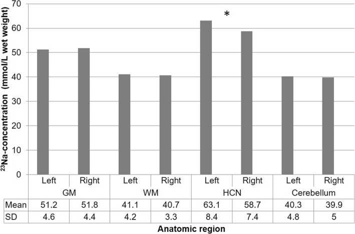
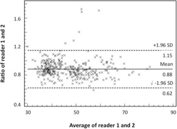
Results: Excellent correlation was found between repeated measurements on the same day (r2 = 0.94), as well as on a different day (r2 = 0.86). No significant differences were found based on laterality other than in the HCN (63.1 vs. 58.7 mmol/kg WW on the right (p = 0.01)). Pronounced inter-individual differences were identified in all anatomic regions. Moderate to good correlation (0.310 to 0.701) was found between the readers.
Conclusion: Our study has shown that intra-individual 23Na-concentrations in healthy subjects do not significantly differ after repeated scans on the same day and a pre-set time interval. This confirms the repeatability and reproducibility of cerebral 23Na-imaging. However, with manual ROI placement in predetermined anatomic landmarks, fluctuations in 23Na-concentrations can be observed.
Keywords: 23Na (sodium) imaging; Cerebral magnetic resonance imaging; Oncologic imaging.
Conflict of interest statement
Ethics approval and consent to participate
The local institutional review board (IRB) and ethics committee (Medical Ethics Committee II Mannheim, Germany; reference number: 2013 566 N – MA) approved this prospective baseline study which was performed in accordance with the ethical standards laid down in the 1964 Declaration of Helsinki and its later amendments.
After the procedure was fully explained, written informed consent was obtained from all participants prior to MR imaging.
Consent for publication
Not applicable.
Competing interests
The authors declare that they have no competing interests.
Publisher’s Note
Springer Nature remains neutral with regard to jurisdictional claims in published maps and institutional affiliations.
Figures


Similar articles
-
Cerebral sodium (23Na) magnetic resonance imaging in patients with migraine - a case-control study.Eur Radiol. 2019 Dec;29(12):7055-7062. doi: 10.1007/s00330-019-06299-1. Epub 2019 Jul 1. Eur Radiol. 2019. PMID: 31264011 Free PMC article.
-
Measuring tissue sodium concentration: Cross-vendor repeatability and reproducibility of 23 Na-MRI across two sites.J Magn Reson Imaging. 2019 Oct;50(4):1278-1284. doi: 10.1002/jmri.26705. Epub 2019 Mar 12. J Magn Reson Imaging. 2019. PMID: 30859655 Free PMC article.
-
Repeatability of Brain Volume Measurements Made with the Atlas-based Method from T1-weighted Images Acquired Using a 0.4 Tesla Low Field MR Scanner.Magn Reson Med Sci. 2016 Oct 11;15(4):365-370. doi: 10.2463/mrms.mp.2015-0107. Epub 2016 Feb 3. Magn Reson Med Sci. 2016. PMID: 26841856 Free PMC article.
-
Brain Magnetic Resonance Imaging Findings in Children and Young Adults With CKD.Am J Kidney Dis. 2018 Sep;72(3):349-359. doi: 10.1053/j.ajkd.2017.11.024. Epub 2018 Feb 15. Am J Kidney Dis. 2018. PMID: 29398180 Free PMC article.
-
Recent technical developments and clinical research applications of sodium (23Na) MRI.Prog Nucl Magn Reson Spectrosc. 2023 Nov-Dec;138-139:1-51. doi: 10.1016/j.pnmrs.2023.04.002. Epub 2023 Apr 18. Prog Nucl Magn Reson Spectrosc. 2023. PMID: 38065665 Review.
Cited by
-
Evaluation of Sodium (23Na) MR-imaging as a Biomarker and Predictor for Neurodegenerative Changes in Patients With Alzheimer's Disease.In Vivo. 2021 Jan-Feb;35(1):429-435. doi: 10.21873/invivo.12275. In Vivo. 2021. PMID: 33402493 Free PMC article.
-
Frontiers of Sodium MRI Revisited: From Cartilage to Brain Imaging.J Magn Reson Imaging. 2021 Jul;54(1):58-75. doi: 10.1002/jmri.27326. Epub 2020 Aug 26. J Magn Reson Imaging. 2021. PMID: 32851736 Free PMC article. Review.
-
23 Na-MRI as a Noninvasive Biomarker for Cancer Diagnosis and Prognosis.J Magn Reson Imaging. 2021 Apr;53(4):995-1014. doi: 10.1002/jmri.27147. Epub 2020 Mar 26. J Magn Reson Imaging. 2021. PMID: 32219933 Free PMC article. Review.
-
Quantitative Sodium (23Na) MRI in Pediatric Gliomas: Initial Experience.Diagnostics (Basel). 2022 May 13;12(5):1223. doi: 10.3390/diagnostics12051223. Diagnostics (Basel). 2022. PMID: 35626378 Free PMC article.
-
The use of 7T MRI in multiple sclerosis: review and consensus statement from the North American Imaging in Multiple Sclerosis Cooperative.Brain Commun. 2024 Oct 9;6(5):fcae359. doi: 10.1093/braincomms/fcae359. eCollection 2024. Brain Commun. 2024. PMID: 39445084 Free PMC article. Review.
References
-
- Haneder S, et al. Quantitative in vivo 23Na MR imaging of the healthy human kidney: determination of physiological ranges at 3.0T with comparison to DWI and BOLD. MAGMA. 2013;26(6):501–9. - PubMed
MeSH terms
Substances
LinkOut - more resources
Full Text Sources
Medical
