. 2019 Apr 3;11(486):eaav3488.
doi: 10.1126/scitranslmed.aav3488.

Mucus accumulation in the lungs precedes structural changes and infection in children with cystic fibrosis

Affiliations

Mucus accumulation in the lungs precedes structural changes and infection in children with cystic fibrosis

Charles R Esther Jr et al. Sci Transl Med. .

Abstract

Although destructive airway disease is evident in young children with cystic fibrosis (CF), little is known about the nature of the early CF lung environment triggering the disease. To elucidate early CF pulmonary pathophysiology, we performed mucus, inflammation, metabolomic, and microbiome analyses on bronchoalveolar lavage fluid (BALF) from 46 preschool children with CF enrolled in the Australian Respiratory Early Surveillance Team for Cystic Fibrosis (AREST CF) program and 16 non-CF disease controls. Total airway mucins were elevated in CF compared to non-CF BALF irrespective of infection, and higher densities of mucus flakes containing mucin 5B and mucin 5AC were observed in samples from CF patients. Total mucins and mucus flakes correlated with inflammation, hypoxia, and oxidative stress. Many CF BALFs appeared sterile by culture and molecular analyses, whereas other samples exhibiting bacterial taxa associated with the oral cavity. Children without computed tomography-defined structural lung disease exhibited elevated BALF mucus flakes and neutrophils, but little/no bacterial infection. Although CF mucus flakes appeared "permanent" because they did not dissolve in dilute BALF matrix, they could be solubilized by a previously unidentified reducing agent (P2062), but not N-acetylcysteine or deoxyribonuclease. These findings indicate that early CF lung disease is characterized by an increased mucus burden and inflammatory markers without infection or structural lung disease and suggest that mucolytic and anti-inflammatory agents should be explored as preventive therapy.


Figures

Figure 1.
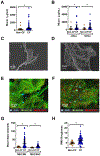
Figure 1.. Mucins in CF and non-CF disease controls.
A) BALF total mucin concentrations from patients with Subjects with CF (n=121) and non-CF controls (n=27). B) Total concentrations of mucins in BALF after stratification of patients by the absence (n=14 non-CF, 82 CF, left) or presence (n=13 non-CF, 39 CF, right) of pathogenic infection (infxn) on culture of BALF. C) Representative SEM of a flake from a non-CF subject. D) Representative SEM of a flake from a CF subject. E. F) Representative IHC of MUC5B (green) and MUC5AC (red) in a cytospin from (E) a non-CF and (F) a CF BALF G) Mucin staining intensities, an index of flake number, of MUC5B and MUC5AC in CF (n=109) and non-CF (n=21) BALF. H) Flake granularity, measured via image analysis of root mean squared roughness in CF ( n=17) and non-CF (n=59) BALF. *p<0.05; ** p<0.01 after multivariate analysis.
Figure 2.
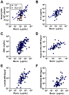
Figure 2.. Mucins and markers of airways disease in early CF.
A) Correlation between mucins and neutrophil counts in BALF from subjects with CF (blue, r2=0.38, p<0.001, n=62) and non-CF controls (orange, n=16, not significant). B) Correlations between mucins and other markers associated with neutrophilic inflammation including IL-8 (r2=0.29, p<0.001, n=62) and C) DNA (r2=0.43, p<0.001, n=62). D-F). Correlations between mucins and BALF metabolite concentrations measured by metabolomics in 60 CF BALF samples, including D) hypoxanthine (r2=0.47, p<0.001) as a marker of inflammation, E) lactate (r2=0.48, p<0.001) as a marker of hypoxia and F) ratio of oxidized to reduced glutathione (GSSG/GSH) as a marker of oxidative stress (r2=0.33, p<0.001).
Figure 3.
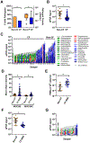
Figure 3.. Bacterial cultures and the microbiome in early CF.
A) Pathogen recovery frequency (left) and burden of pathogens in samples with positive cultures (right) in BALF from subjects with CF and from non-CF controls (n=124 CF, 32 non-CF). B) Total bacterial burden, as measured by 16S qPCR, in BALF from subjects with CF and non-CF controls (n=46 CF, 15 non-CF). C) qPCR and distribution of taxa from microbiome analyses for all CF (left) and non-CF samples (right). Bar height represents total qPCR signal, with detected taxa represented proportionally within the bar. For clarity, taxa with average prevalence <1% were grouped (light gray). The pattern observed from washes of sterile bronchoscopes (w) is shown for reference (n=6). Taxa associated with environmental contamination are shown in green, taxa associated with the oral cavity, including Moraxella, Haemophilus, and Streptococcus as common upper airway commensals (72), are shown in blue, and known pathogens are shown in red. Taxa were grouped as previously described (73) D) Concentrations of MUC5B and MUC5AC as measured by IHC in mucin flakes recovered by BALF from subjects with CF with no structural lung disease (CF-NSD, n=37) and non-CF controls (n=21) (significant by parameter; p=0.054 post-multivariate analyses). E) Neutrophil (PMN) counts in CF-NSD vs. non-CF. F) qPCR in CF-NSD and non-CF samples. G) Microbiome analyses of CF-NSD. Triangles represent samples from subjects who received antibiotics ≤3 months prior to bronchoscopy. See panel 3C for taxa legend. *=p<0.05, †=p<0.05 by non-parametric analysis.
Figure 4.
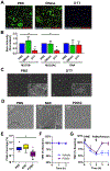
Figure 4.. Treatment of mucin flakes in early CF.
A) IHC of MUC5B (green), MUC5AC (red), and DNA (Blue) on a cytospin of unprocessed CF BALF treated with (PBS), DNase (300 units/ml), or the reducing agent DTT (10 mM) for 60 minutes at 37ºC. B) Staining intensities of MUC5B and MUC5AC in DTT treated samples relative to those with vehicle (PBS) or DNase (n=5/group). Lack of effect of DNase on DNA staining intensities reflected the intracellular location of most DNA. All intensities were normalized to the average values from the PBS group (n=10/group). C) Flake density in PBS and DTT treated samples analyzed by SEM. D) Flake density and structure by SEM in samples treated with NAC (10 mM, 1 hour at 37º) or the novel therapeutic P2062 (10 mM, 1 hour at 37º). E) Imaging analysis of flake density in CF BALF samples treated with NAC or P2062 (n=7/group). F) Tracheal mean velocity (TMV), a measure of mucociliary clearance, in sheep treated with nebulized P2062 (2.5 mL. 50 mM concentration, solid purple line) or saline (dotted blue line) (n=3/group). G) TMV in sheep exposed to inhaled neutrophil elastase followed by treatment with nebulized P2062 (solid purple line) or saline (dotted blue line) did not (n=3/group).

References

    1. Davis PB, Cystic Fibrosis Since 1938. Am. J. Respir. Crit. Care Med 173, 475–482 (2006). - PubMed
    1. Mott LS et al., Progression of early structural lung disease in young children with cystic fibrosis assessed using CT. Thorax 67, 509–516 (2012). - PubMed
    1. Sly PD et al., Lung disease at diagnosis in infants with cystic fibrosis detected by newborn screening. Am J Respir Crit Care Med 180, 146–152 (2009). - PubMed
    1. Ehre C, Ridley C, Thornton DJ, Cystic fibrosis: an inherited disease affecting mucin-producing organs. The international journal of biochemistry & cell biology 52, 136–145 (2014). - PMC - PubMed
    1. Farber S, Shwachman H, Maddock CL, Pancreatic Function and Disease in Early Life. I. Pancreatic Enzyme Activity and the Celiac Syndrome. J Clin Invest 22, 827–838 (1943). - PMC - PubMed

Publication types