. 2019 Apr 4;4(7):e123253.
doi: 10.1172/jci.insight.123253.

Vimentin intermediate filament assembly regulates fibroblast invasion in fibrogenic lung injury

Affiliations

Vimentin intermediate filament assembly regulates fibroblast invasion in fibrogenic lung injury

Ranu Surolia et al. JCI Insight. .

Abstract

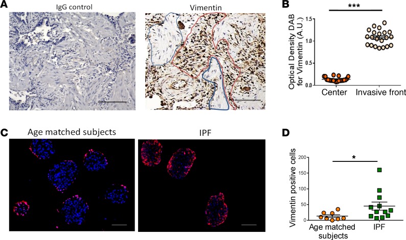
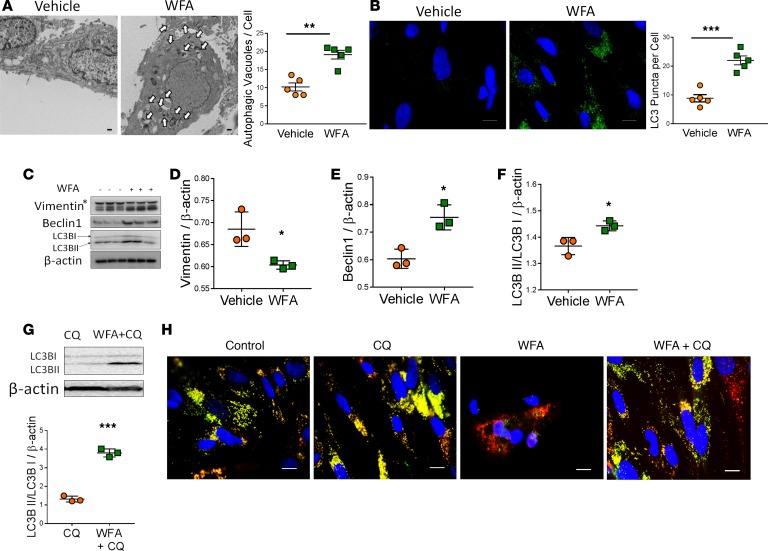
Idiopathic pulmonary fibrosis (IPF) is a progressive disease, with a median survival of 3-5 years following diagnosis. Lung remodeling by invasive fibroblasts is a hallmark of IPF. In this study, we demonstrate that inhibition of vimentin intermediate filaments (VimIFs) decreases the invasiveness of IPF fibroblasts and confers protection against fibrosis in a murine model of experimental lung injury. Increased expression and organization of VimIFs contribute to the invasive property of IPF fibroblasts in connection with deficient cellular autophagy. Blocking VimIF assembly by pharmacologic and genetic means also increases autophagic clearance of collagen type I. Furthermore, inhibition of expression of collagen type I by siRNA decreased invasiveness of fibroblasts. In a bleomycin injury model, enhancing autophagy in fibroblasts by an inhibitor of VimIF assembly, withaferin A (WFA), protected from fibrotic lung injury. Additionally, in 3D lung organoids, or pulmospheres, from patients with IPF, WFA reduced the invasiveness of lung fibroblasts in the majority of subjects tested. These studies provide insights into the functional role of vimentin, which regulates autophagy and restricts the invasiveness of lung fibroblasts.

Keywords: Autophagy; Drug therapy; Fibrosis; Pulmonology.


Conflict of interest statement

Conflict of interest: The authors have declared no conflict of interest.

Figures

Figure 1
Figure 1. Vimentin expression is upregulated at the margins of IPF fibrotic foci.
(A) IHC staining showing IgG control and expression of vimentin in formalin-fixed, paraffin-embedded tissue section of a representative subject with IPF. Scale bars: 100 μm. Area outlined in red is the invasive front of fibrotic foci (outlined in blue). (B) Quantification of DAB-positive vimentin-expressing cells in the center and at the cellular cuffs of fibrotic foci. Graph shows representative results for an individual subject (n = 5). Each dot represents DAB intensity per area. Data are represented as mean ± SD. (C) Representative image of the expression of vimentin in pulmospheres obtained from the cells isolated from control and IPF lung tissue. The pulmospheres were seeded for an hour in collagen gel prior to immunostaining for vimentin (red). Nuclei were stained with DAPI (blue). Scale bars: 100 μm. (D) Quantification of vimentin-positive cells in pulmospheres from controls and IPF subjects. Data are represented as mean ± SD. Each dot represents an individual subject. *P < 0.05, ***P < 0.001.
Figure 2
Figure 2. Vimentin expression is upregulated during attenuated autophagy in IPF fibroblasts.
(A) Immunoblot analysis of vimentin and the autophagic markers beclin 1 and LC3BII in cell lysates of fibroblasts from age-matched subjects (n = 5) and IPF (n = 5) lung tissue in complete medium (control) and serum-starved conditions. β-Actin served as loading control. Densitometry analysis of protein expression in Western blot for (B) vimentin, (C) beclin 1, and (D) LC3BII/LC3BI normalized with β-actin in controls and IPF fibroblasts. *P < 0.05. (E) Invasiveness in the pulmospheres obtained from age-matched subjects and IPF lung in complete medium and serum-starved conditions. Data are presented as mean ± SD; each dot represents an individual patient. **P < 0.01. Vimentin expression in complete medium and serum-starved conditions in pulmospheres from age-matched subjects and IPF patients (F) (scale bars: 200 μm); quantitation by FACS analysis of vimentin-positive cells in dissociated pulmospheres (G). (H) Single invasive fibroblast in pulmospheres (scale bars: 10 μm).
Figure 3
Figure 3. Inhibition of VimIF assembly induces autophagy in IPF fibroblasts.
(A) Representative electron photomicrograph and quantitative graph showing increased number of autophagic vacuoles in vehicle and WFA-treated IPF fibroblasts. Data are presented as mean ± SEM. Three cells per IPF patient were analyzed for the quantification of autophagic vacuoles. Each dot represents an individual patient. **P < 0.01, as compared with vehicle-treated fibroblasts. Scale bars: 10 μm. (B) Human fibroblasts were treated with vehicle or WFA, and laser confocal microscopy was performed to detect LC3B by indirect immunofluorescence. Nuclei were costained with DAPI (blue). Scale bars: 20 μm. Graph represents the quantification of LC3BII puncta in WFA-treated fibroblasts. ***P < 0.001, as compared with vehicle-treated fibroblasts. Data are mean ± SD (n = 5 patients). (C) Immunoblot analysis for vimentin, LC3B, and beclin 1 of cell lysates from WFA-treated IPF fibroblasts. Asterisk indicates vimentin 56-kDa band. Each lane represents an individual subject. β-Actin served as loading control. Densitometry for (D) vimentin (band at 56 kDa), (E) beclin 1, and (F) LC3BII/LC3B I ratio normalized to β-actin for the Western blot. *P < 0.05, compared with vehicle-treated fibroblasts. (G) Immunoblot analysis for LC3B of cell lysates from IPF fibroblasts treated with chloroquine (CQ) and cotreated with WFA/CQ. β-Actin served as loading control. Data are representative of 3 experiments. The graph represents densitometry analysis for LC3BII/LC3B I ratio normalized to β-actin for the immunoblot blot analysis of LC3B. ***P < 0.001, compared with vehicle-treated fibroblasts. (H) Representative images of cells expressing the RFP-GFP-LC3B construct followed by the treatment with vehicle alone (control) or CQ, WFA, and WFA/CQ cotreatment for 12 hours. CQ-treated cells served as a positive control. Nuclei were costained with DAPI (blue). Natural-pH LC3B-positive autophagosome (green fluorescence) and acidic-pH LC3B-positive autophagolysosome (red fluorescence) were detected respectively using a fluorescence microscope. Scale bars: 20 μm
Figure 4
Figure 4. WFA-regulated autophagy is beclin 1 dependent.
(A) Immunoblot analysis and densitometry for beclin 1 and LC3B of cell lysates from nontargeted siRNA– and beclin 1 siRNA–transfected (siNT- and siBeclin1-transfected) IPF fibroblasts treated with WFA. β-Actin served as loading control. Data are representative of 3 experiments. (B) Immunoblot analysis and densitometry of collagen I (Col1), vimentin, and beclin 1 from cell lysates from siNT- and siBeclin1-transfected IPF fibroblasts treated with WFA. β-Actin served as loading control. Data are representative of 3 experiments. *P < 0.05, **P < 0.01, ***P < 0.001. (C) Representative image and measurement of invasiveness (percent zone of invasion) in beclin 1 siRNA–transfected IPF pulmospheres. siNT was used as control. Scale bars: 100 μm. **P < 0.01, compared with siNT fibroblasts. (D) Immunoprecipitation studies with anti–beclin 1 antibodies of cell lysates from IPF fibroblasts treated with vehicle and WFA. The graph represents vimentin/beclin 1 binding ratio. Precipitates were immunoblotted for associated beclin 1 and vimentin, and whole cell lysates (WCL) for LC3B, vimentin, and beclin 1 antibodies. β-Actin served as loading control for cell lysates. (E) Immunostaining for beclin 1 (green) and vimentin (red) in IPF fibroblasts treated in the absence or presence of WFA. Scale bars: 20 μm.
Figure 5
Figure 5. Inhibition of VimIF assembly induces autophagic clearance of collagen type I in IPF fibroblasts.
(A) Immunoblot analysis for vimentin and collagen type I (Col I) on cell lysates of IPF fibroblasts treated with WFA for different durations. β-Actin served as loading control. Data are representative of 3 experiments. (B) Immunostaining for Col I in IPF fibroblasts treated with vehicle (control) or WFA. Nuclei were costained with DAPI. Scale bars: 20 μm. (C) IPF fibroblasts were treated with vehicle (control) or WFA, and the presence of Col I in autophagosomes was assessed by indirect immunofluorescence and visualization of Col I– and LC3B-positive structures by immunofluorescence microscopy. Scale bars: 20 μm. Inset box shows magnified area of interest. (D) Measurement of invasiveness (percent zone of invasion) in IPF patient pulmospheres after treatment with an autophagy inhibitor (chloroquine [CQ]) or autophagy inducer (rapamycin [Rapa.]). Each dot represents an individual patient. *P < 0.05, **P < 0.01, as compared with control. (E and F) Immunoblot of Col I siRNA– transfected (siCOL1) human fibroblasts and measurement of invasiveness (zone of invasion percent) in IPF pulmospheres after siCOL1 transfection in the presence or absence of WFA. **P < 0.005, ***P < 0.001. (G) Fibroblasts from IPF subjects were treated with WFA to evaluate the expression of Col I. β-Actin served as loading control. (H) Quantification of soluble collagen in the supernatants of IPF fibroblasts treated with vehicle (control) or WFA. *P < 0.05, compared with vehicle-treated fibroblasts.
Figure 6
Figure 6. WFA protects from pulmonary fibrosis in bleomycin mice model.
(A) Schematic of animal model and WFA treatment (B) LC3B II puncta formation, as assessed by immunofluorescence staining for LC3B (green) and nuclei (DAPI, blue) in paraffin embedded mouse lung sections on day 21. Scale bar: 100 μm. Bleo., bleomycin. (C) Quantification of intensity of LC3BII puncta in immunofluorescently labeled mouse lung sections by ImageJ. (D) Immunoblot analysis and (E) densitometry for beclin 1, p62, vimentin, and LC3B in whole lung homogenates from C57BL/6 mice subjected to control saline, bleomycin, WFA, and bleomycin and WFA treatment on day 21. β-Actin served as loading control. Data are representative of 3 experiments. (F) Representative images and measurement of invasiveness (zone of invasion percent) in pulmospheres prepared from lung cells from C57BL/6 mice subjected to control saline, bleomycin, WFA and bleomycin/WFA on day 21. Scale bars: 100 μm. (G) Representative images of immunofluorescence staining for vimentin (green), LC3B (red), and nucleus (DAPI, blue) in pulmospheres prepared from mouse lungs cells after 21 days of bleomycin and bleomycin/WFA exposure. Scale bar: 50 μm (H) MicroCT analysis of mouse lungs on day 20. (I) Fibrosis, as assessed on day 21 by H&E staining. Collagen deposition was analyzed using Picrosirius staining and IHC analysis of Col 1. Scale bar: 100 μm. (J) Lung homogenates analyzed on day 21 for hydroxyproline content. Values represent means ±SD, n = 5. *P < 0.05, **P < 0.01, ***P < 0.001.
Figure 7
Figure 7. Induction of autophagy by inhibition of VimIF assembly decreases invasiveness.
Stable transfection with mEmerald-vimentin construct (WT) and GFP-vimentin-C328S construct (C328S) in Vim–/– mouse fibroblasts. (A) Fluorescence images showing arrangement of vimentin in WT and C328S fibroblasts. Scale bars: 50 μm. (B) Immunoblot analysis for LC3B and vimentin in transfected fibroblasts after 6 hours of serum starvation. β-Actin served as loading control. Densitometry data are representative of 3 experiments. (C) Immunofluorescence staining of pulmospheres using WT and C328 fibroblasts for LC3B (red) and green fluorescence–tagged vimentin expression after 6 hours of serum starvation. Scale bars: 100 μm. (D) Fluorescence images of IPF pulmospheres containing mEmerald-vimentin–transfected IPF fibroblasts treated with vehicle (control) or WFA. Scale bars: 250 μm. (E) Representative image of pulmospheres treated with vehicle or WFA. Graph shows calculated zone of invasion percentage from 6 IPF lung pulmospheres from controls not treated and treated with WFA. Each dot represents the mean value of calculated zone of invasion from 5 pulmospheres for each IPF subject. Data are expressed as mean ± SEM. ***P = 0.002, compared with vehicle-treated pulmospheres. (F) Transmission electron microscopy images of vehicle- and WFA-treated pulmospheres. Scale bars: 2 μm; magnified images, 500 nm. (G) Calculation formula for determination of fold change ratio for zone of invasion (ZOI) area. Median values of ZOI ≥1 represent pulmospheres nonresponsive to the treatment with WFA, and median values of ZOI <1 represent pulmospheres responsive to WFA treatment. (H) Invasiveness of pulmospheres from an individual patient not treated and treated with WFA. Box-and-whisker plots for IPF patient pulmospheres treated with WFA. Whiskers show maximum to minimum values. Lines in boxes represent median values.

References

    1. Ahluwalia N, Shea BS, Tager AM. New therapeutic targets in idiopathic pulmonary fibrosis. Aiming to rein in runaway wound-healing responses. Am J Respir Crit Care Med. 2014;190(8):867–878. - PMC - PubMed
    1. Wolters PJ, Collard HR, Jones KD. Pathogenesis of idiopathic pulmonary fibrosis. Annu Rev Pathol. 2014;9:157–179. - PMC - PubMed
    1. Basset F, Ferrans VJ, Soler P, Takemura T, Fukuda Y, Crystal RG. Intraluminal fibrosis in interstitial lung disorders. Am J Pathol. 1986;122(3):443–461. - PMC - PubMed
    1. White ES, et al. Integrin alpha4beta1 regulates migration across basement membranes by lung fibroblasts: a role for phosphatase and tensin homologue deleted on chromosome 10. Am J Respir Crit Care Med. 2003;168(4):436–442. - PMC - PubMed
    1. Torry DJ, Richards CD, Podor TJ, Gauldie J. Anchorage-independent colony growth of pulmonary fibroblasts derived from fibrotic human lung tissue. J Clin Invest. 1994;93(4):1525–1532. - PMC - PubMed

Publication types

MeSH terms