. 2019 May;17(5):4094-4101.
doi: 10.3892/ol.2019.10079. Epub 2019 Feb 27.

Non-invasive imaging techniques for the in vivo diagnosis of Bowen's disease: Three case reports

Affiliations

Non-invasive imaging techniques for the in vivo diagnosis of Bowen's disease: Three case reports

Simona Laura Ianoși et al. Oncol Lett. 2019 May.

Abstract

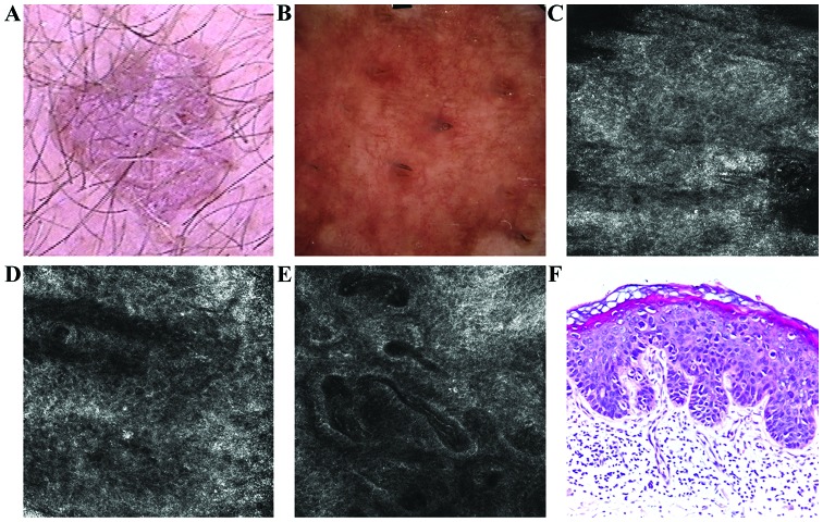
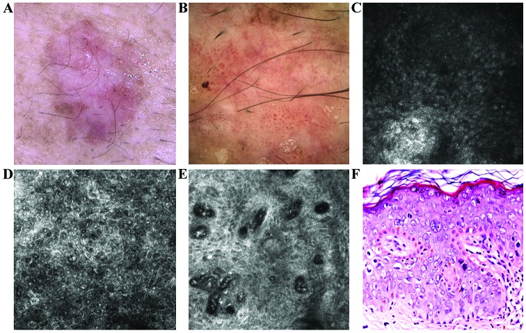
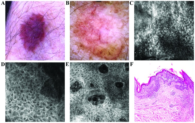
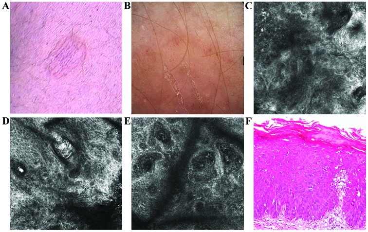
Bowen's disease (BD) is a relatively frequent non-melanoma skin cancer occurring mostly in elderly people. Until now, the usual way to establish the diagnosis is histopathological examination of a skin biopsy. Dermoscopy and reflectance confocal microscopy (RCM) are modern alternative methods that can be used as quick and non-invasive diagnostic techniques and as follow-up instruments in cases in which a conservative treatment is chosen for the management of BD. There are no very specific dermoscopic criteria for the diagnosis of this disease, but some dermoscopic features (scaly surface, vascular structures and pigmentation) can be found more frequent and can be helpful for the diagnosis. RCM of BD shows an acanthotic epidermis with two types of targetoid cells: the first, a large cell with bright center and dark peripheral halo, the second, a cell with dark center and a bright rim surrounded by a dark hallo, related with dyskeratotic cells on histological examination. BD management could be improved by using non-invasive, in vivo imaging techniques that allow a fast and easy diagnosis and can be used as follow-up tools. However, larger studies are necessary for the validation of our observations.

Keywords: Bowen's disease; dermoscopy; diagnosis; in vivo; non-invasive; reflectance confocal microscopy.


Figures

Figure 1.
Figure 1.
(A) Clinical image illustrated a well-demarcated, erythematous, scaly plaque. (B) Dermoscopic aspect of the lesion revealed vascular pattern with dotted, linear, comma-like and glomerular vessels. (C) RCM examination (500×500 µm) of the stratum corneum showed the presence of bright, polygonal shaped structures corresponding to parakeratotic cells (D) RCM image (500×500 µm) with chaotic appearance of the spinous layer resulted in an atypical honeycomb pattern and sparse target-like cells. (E) RCM image (500×500 µm) of the dermal-epidermal junction revealed enlarged, irregular shaped papillae filled with dilated capillaries, with reduction of the papillary rings normally seen in healthy skin. (F) Histopathological image showed areas of parakeratosis, numerous dyskeratotic keratinocytes, moderate cellular and nuclear pleomorphism and numerous mitosis; moderate hyperemia and lymphocytic inflammatory infiltrate (hematoxylin and eosin staining; ×200 magnification).
Figure 2.
Figure 2.
(A) Clinical image of a flat plaque showed pink and brown diffuse areas. (B) Corresponding dermoscopic image showed diffuse, structureless brown pigmentation, some brown globules and an atypical vascular pattern consist of glomerular, dotted and linear vessels. (C) RCM image (500×500 µm) at the level of the stratum corneum where parakeratotic cells were identified as round to polygonal, refractile structures. (D) RCM image (500×500 µm) revealed the atypical honeycomb pattern in the spinous layer, with various shapes and sizes of the cells and nuclei. Many of these cells have a target-like aspect, with a bright center and a dark peripheral halo. Another type of cell with targetoid aspect, with a dark center and a bright peripheral rim, was also identified in smaller numbers. (E) RCM image (500×500 µm) at the level of the dermo-epidermal junction showed enlarged dermal papillae, with bizarre shapes, without the papillary rings of basal cells, inside of which are identified dilated capillaries, some captured in horizontal section, parallel to the skin. (F) Histopathological image displaying important cytonuclear pleomorphism with hypertrophy and nuclear monstrosities and atypical mitosis at different levels in epidermal thickness (hematoxylin and eosin staining; ×200 magnification).
Figure 3.
Figure 3.
(A) Clinical image of a slightly variegated brown plaque with very fine scaling. (B) Dermoscopic view showed structureless asymmetric brown pigmentation with erythematous background, few brown globules and irregular network in the periphery and an atypical vascular pattern. (C) RCM examination (500×500 µm) revealed parakeratosis in the horny layer. (D) RCM image (500×500 µm) at the level of the spinous-granular layers showed a disarrayed pattern and target-like cells. (E) RCM image (500×500 µm) at the dermo-epidermal junction the dermal papillae are filled with many dilated capillaries. (F) Corresponding histology with epidermal thickening due to proliferation of atypical squamous cells with cyto-nuclear atypia extended through the whole thickness of the epidermis (hematoxylin and eosin staining; ×100 magnification).
Figure 4.
Figure 4.
(A) Clinical image showed a scaly, pink and light brown, flat plaque, with well-defined borders. (B) Dermoscopic image showed coil-shaped and dotted vessels and light brown structureless areas on a pink background. (C) RCM image (500×500 µm) revealed a parakeratosis area in the horny layer (upper left). (D) RCM image (500×500 µm) at the level of the spinous layer showing an atypical honeycomb pattern. (E) RCM image (500×500 µm) at the dermo-epidermal junction revealing enlarged dermal papillae with dilated and sinuous blood vessels. (F) Histopathological image showing epidermal thickening with areas of parakeratosis, atypical keratinocytes, moderate pleomorphism and moderate lymphocytic inflammatory infiltrate (hematoxylin and eosin staining; ×200 magnification).

References

    1. Cox NH, Eedy DJ, Morton CA, British Association of Dermatologists Guidelines for management of Bowen's disease. Br J Dermatol. 1999;141:633–641. doi: 10.1046/j.1365-2133.1999.03100.x. - DOI - PubMed
    1. Cox NH, Eedy DJ, Morton CA, Therapy Guidelines and Audit Subcommittee, British Association of Dermatologists Guidelines for management of Bowen's disease: 2006 update. Br J Dermatol. 2007;156:11–21. doi: 10.1111/j.1365-2133.2006.07610.x. - DOI - PubMed
    1. Weinberg AS, Ogle CA, Shim EK. Metastatic cutaneous squamous cell carcinoma: An update. Dermatol Surg. 2007;33:885–899. doi: 10.1111/j.1524-4725.2007.33190.x. - DOI - PubMed
    1. Krishnan R, Lewis A, Orengo IF, Rosen T. Pigmented Bowen's disease (squamous cell carcinoma in situ): A mimic of malignant melanoma. Dermatol Surg. 2001;27:673–674. doi: 10.1046/j.1524-4725.2001.01021.x. - DOI - PubMed
    1. Cameron A, Rosendahl C, Tschandl P, Riedl E, Kittler H. Dermatoscopy of pigmented Bowen's disease. J Am Acad Dermatol. 2010;62:597–604. doi: 10.1016/j.jaad.2009.06.008. - DOI - PubMed