Review
. 2019 Jun;20(6):404-435.
doi: 10.1111/tra.12646.

The road to lysosome-related organelles: Insights from Hermansky-Pudlak syndrome and other rare diseases

Affiliations
Review

The road to lysosome-related organelles: Insights from Hermansky-Pudlak syndrome and other rare diseases

Shanna L Bowman et al. Traffic. 2019 Jun.

Abstract

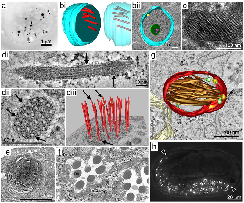
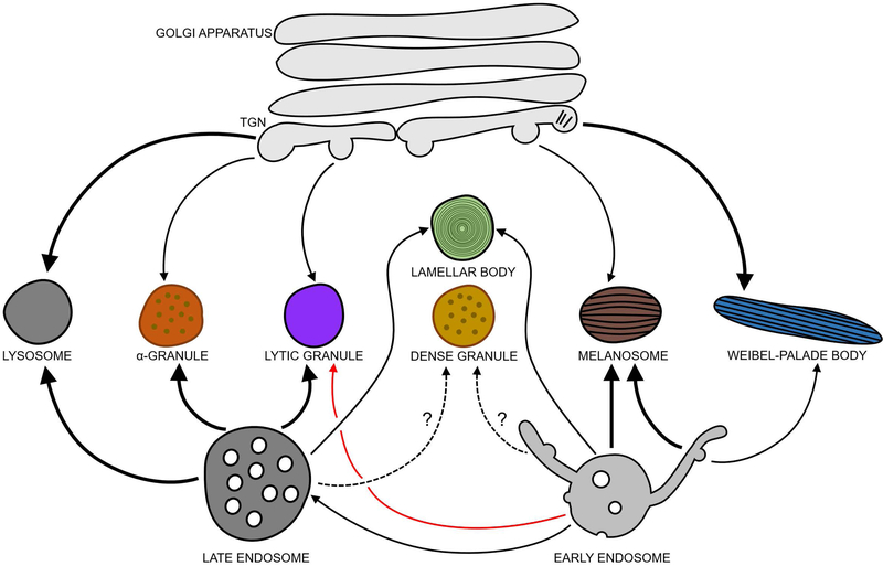
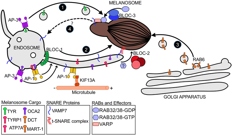
Lysosome-related organelles (LROs) comprise a diverse group of cell type-specific, membrane-bound subcellular organelles that derive at least in part from the endolysosomal system but that have unique contents, morphologies and functions to support specific physiological roles. They include: melanosomes that provide pigment to our eyes and skin; alpha and dense granules in platelets, and lytic granules in cytotoxic T cells and natural killer cells, which release effectors to regulate hemostasis and immunity; and distinct classes of lamellar bodies in lung epithelial cells and keratinocytes that support lung plasticity and skin lubrication. The formation, maturation and/or secretion of subsets of LROs are dysfunctional or entirely absent in a number of hereditary syndromic disorders, including in particular the Hermansky-Pudlak syndromes. This review provides a comprehensive overview of LROs in humans and model organisms and presents our current understanding of how the products of genes that are defective in heritable diseases impact their formation, motility and ultimate secretion.

Keywords: AP-3; BLOC-1; BLOC-2; BLOC-3; Chediak-Higashi syndrome; Griscelli syndrome; HOPS; Hermansky-Pudlak syndrome; RAB27A; RAB32; RAB38; VPS33A; VPS33B; Weibel-Palade body; alpha granule; dense granule; lamellar body; melanosome.


Figures

Figure 1:
Figure 1:. Examples of lysosome-related organelles.
(a) Dense granules (arrows) in a human platelet observed by whole mount electron microscopy from ref. 447. Scale bar, 1 μm. (bi – bii) 3D reconstructions of α-granules in chemically fixed human platelets analyzed by electron tomography from ref. 448. The limiting membrane is in blue. Scale bar, 50 nm. (bi) Transverse view (left) and side view with transparent membrane (right) emphasizing the arrangement of VWF tubules (red on left, red-gray on right) within the organelle. (bii) Transverse view of an alpha granule that is immunogold labeled for P-selectin (yellow) and CD63 (magenta), emphasizing an intraluminal vesicle (green). (c) A lamellar body from ex vivo human skin analyzed by thin section electron microscopy (from ref. 449). Scale bar, 100 nm. (di – diii) Electron tomography of WPBs in human umbilical vein endothelial cells from ref. 450. (di) A longitudinal tomographic slice of a WPB emphasizing vWF tubules (arrows) along the length. (dii) A transverse tomographic section of a WPB emphasizing the diameter of vWF tubules. (diii) 3D reconstruction of (dii) showing the extension of individual VWF tubules. Arrows point to VWF tubules that end halfway through the WPB. Scale bars, 100 nm. (e) A lung lamellar body from a rat type II alveolar cell analyzed by thin section electron microscopy (from ref. 451). Scale bar, 1 μm. (f) Transverse section of a wild-type D. melanogaster eye analyzed by thin section electron microscopy from ref. 77. Note the pigment granules (PG) of secondary pigment cells surrounding photoreceptor cell rhabdomeres (Rh). (g) 3D reconstruction of a stage II melanosome from electron tomography analysis of a human MNT-1 melanoma cell (from ref. 452). Red, melanosome membrane; brown, intraluminal fibrils; intralumenal vesicles are in yellow (membrane-associated) or green (free). Scale bar, 200 nm. (h) Birefringent material in gut granules (arrowheads) in a C. elegans embryo observed by polarization microscopy (from ref. 279). Scale bar, 20 μm. All panels reprinted by permission of: (a) Taylor and Francis from Platelets, ref. 447, copyright 2018; (b) American Society of Hematology from Blood, ref. 448, copyright 2010; (c) Elsevier from the Journal of Dermatological Science, ref. 449, copyright 2018; (d) Elsevier from the Journal of Structural Biology, ref. 450, copyright 2008; (e) Springer Nature from Pediatric Research, ref. 451, copyright 2012; (f) John Wiley and Sons from EMBO Journal, ref. ,, copyright 1997; (g) National Academy of Sciences, U.S.A from Proceedings of the National Academy of Sciences U.S.A., ref. 452, copyright 2008; and (h) PLoS Genetics, ref. 279.
Figure 2.
Figure 2.. Biogenesis models for mammalian LROs.
Lytic granules in CTLs and natural killer cells may derive from late endosomes that fuse with secretory vesicles from the TGN, and also function as the cells’ lysosomes under basal conditions. Lytic granule maturation (by fusion with early endosomal membranes; red arrow) and secretion are triggered by immune stimulation. The other indicated mammalian LROs co-exist with endolysosomes but derive from late endosomes, early endosomes, and/or the TGN as indicated. Question marks denote LRO biogenesis pathways that are not well-characterized, and thick solid lines denote pathways, where known, by which the majority of material is targeted to maturing LROs. Alpha granules obtain material from multivesicular late endosomes in platelets and likely derive from these compartments, but also receive vWF from the TGN. Platelet dense granules and lung AT2 lamellar bodies may derive components from both late endosomes and early endosomes during maturation. In pigment cell melanocytes, immature melanosomes emerge from maturing endosomes and receive transmembrane cargo via early endosomal tubule carriers, endosome-derived vesicles, and the Golgi as they mature. In endothelial cells, immature Weibel-Palade bodies harboring vWF tubules that form in the Golgi bud from the TGN and mature by addition of cargoes derived from endosomes.
Figure 3.
Figure 3.. HPS complexes and mechanisms of cargo delivery to melanosomes.
Melanosome-destined transmembrane protein cargoes are concentrated on early endosomes by AP-3 and AP-1. The majority of TYR (green) transits a vesicular pathway that requires AP-3 (Pathway 1). Other proteins such as TYRP1, OCA2, and ATP7A, transit endosome-derived membrane tubules en route to melanosomes (Pathway 2). BLOC-1 and the KIF13A kinesin motor are required to generate the tubules along microtubules, and KIF13A is recruited to endosomes by AP-1. Cargo sorting on this pathway is mediated by AP-1 and/or AP-3; TYRP1 and ATP7A engage only AP-1, while both AP-1 and AP-3 facilitate OCA2 transport on this pathway. BLOC-2 is required to direct the tubular transport carriers to melanosome membranes, and RAB32 and/or RAB38 might also play a role in this step. Cargo delivery requires transient fusion of the tubular transport carriers with melanosome membranes mediated by the v-SNARE, VAMP7, and an unidentified t-SNARE complex. DCT and MART1 are transported to melanosomes from the Golgi Apparatus in a separate vesicular pathway that requires RAB6 (Pathway 3). VAMP7 and perhaps other cargoes are recycled from melanosomes via tubules that derive from melanosome membranes in a BLOC-3- and RAB38/RAB32-dependent manner (Pathway 4). The scaffolding protein VARP is present on these tubules and likely supports the incorporation of VAMP7 into them. The destination of these tubules is not yet known, but VAMP7 is likely ultimately returned to early endosomes.

References

    1. Huotari J, Helenius A. Endosome maturation. EMBO J. 2011;30(17):3481–3500. - PMC - PubMed
    1. Gould GW, Lippincott-Schwartz J. New roles for endosomes: from vesicular carriers to multi-purpose platforms. Nat Rev Mol Cell Biol. 2009;10(4):287–292. - PMC - PubMed
    1. Bajno L, Peng XR, Schreiber AD, Moore HP, Trimble WS, Grinstein S. Focal exocytosis of VAMP3-containing vesicles at sites of phagosome formation. J Cell Biol. 2000;149(3):697–706. - PMC - PubMed
    1. Collins RF, Schreiber AD, Grinstein S, Trimble WS. Syntaxins 13 and 7 function at distinct steps during phagocytosis. J Immunol. 2002;169(6):3250–3256. - PubMed
    1. Parton RG, Prydz K, Bomsel M, Simons K, Griffiths G. Meeting of the apical and basolateral endocytic pathways of the Madin-Darby canine kidney cell in late endosomes. J Cell Biol. 1989;109(6 part 2):3259–3272. - PMC - PubMed

Publication types

MeSH terms