. 2019 Apr 4;14(4):e0214520.
doi: 10.1371/journal.pone.0214520. eCollection 2019.

Differential changes in bone strength of two inbred mouse strains following administration of a sclerostin-neutralizing antibody during growth

Affiliations

Differential changes in bone strength of two inbred mouse strains following administration of a sclerostin-neutralizing antibody during growth

Noah J Mathis et al. PLoS One. .

Abstract

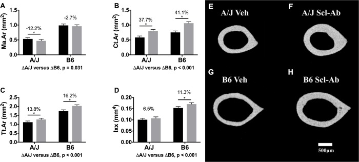
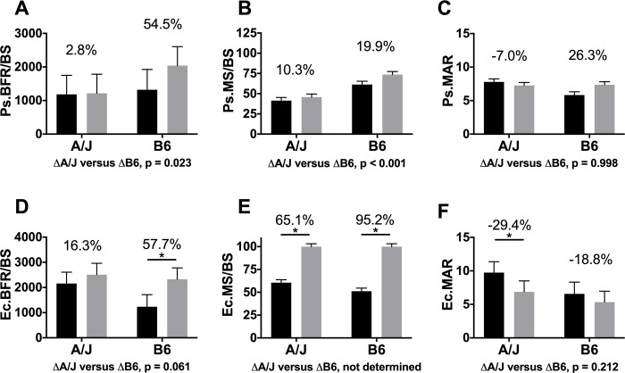
Administration of sclerostin-neutralizing antibody (Scl-Ab) treatment has been shown to elicit an anabolic bone response in growing and adult mice. Prior work characterized the response of individual mouse strains but did not establish whether the impact of Scl-Ab on whole bone strength would vary across different inbred mouse strains. Herein, we tested the hypothesis that two inbred mouse strains (A/J and C57BL/6J (B6)) will show different whole bone strength outcomes following sclerostin-neutralizing antibody (Scl-Ab) treatment during growth (4.5-8.5 weeks of age). Treated B6 femurs showed a significantly greater stiffness (S) (68.8% vs. 46.0%) and maximum load (ML) (84.7% vs. 44.8%) compared to A/J. Although treated A/J and B6 femurs showed greater cortical area (Ct.Ar) similarly relative to their controls (37.7% in A/J and 41.1% in B6), the location of new bone deposition responsible for the greater mass differed between strains and may explain the greater whole bone strength observed in treated B6 mice. A/J femurs showed periosteal expansion and endocortical infilling, while B6 femurs showed periosteal expansion. Post-yield displacement (PYD) was smaller in treated A/J femurs (-61.2%, p < 0.001) resulting in greater brittleness compared to controls; an effect not present in B6 mice. Inter-strain differences in S, ML, and PYD led to divergent changes in work-to-fracture (Work). Work was 27.2% (p = 0.366) lower in treated A/J mice and 66.2% (p < 0.001) greater in treated B6 mice relative to controls. Our data confirmed the anabolic response to Scl-Ab shown by others, and provided evidence suggesting the mechanical benefits of Scl-Ab administration may be modulated by genetic background, with intrinsic growth patterns of these mice guiding the location of new bone deposition. Whether these differential outcomes will persist in adult and elderly mice remains to be determined.


Conflict of interest statement

The authors have declared that no competing interests exist.

Figures

Fig 1
Fig 1. Schematic of key morphological, compositional, and molecular differences between A/J and B6 femurs.
Despite variation in traits, both bones establish similar organ level mechanical function at musculoskeletal maturity. Adapted from Schlecht et al [14].
Fig 2
Fig 2. Representative load-deflection curves from four-point bending.
A) A/J Veh and A/J Scl-Ab, and B) B6 Veh and B6 Scl-Ab. Stiffness was calculated as the slope of the linear portion of the curve, maximum (max) load was calculated as the highest point on the curve, PYD was the displacement between the end of the linear portion of the curve and the point of failure, and work-to-fracture was calculated as area under the curve put to the point of failure.
Fig 3
Fig 3. Four-point bending of the femurs of treated and untreated mice.
Both strains of treated mice showed greater whole bone A) stiffness and B) strength compared to their respective controls. C) PYD was significantly smaller with treatment in A/J but not B6, and D) Work showed divergent outcomes, being significantly smaller in treated A/J femurs but significantly greater in treated B6 femurs compared to their respective controls. Main effects by strain and treatment as well as the interaction of strain by treatment were tested using an ANOVA. Percent changes were calculated from least square mean differences of Tukey pairwise comparisons (p < 0.05). Black bars = Vehicle and gray bars = Scl-Ab treated.
Fig 4
Fig 4. Morphological data derived from the femoral diaphysis.
Data showed that Ct.Ar (A) and Tt.Ar (B) were similarly significantly greater in A/J and B6 femurs, while Ma.Ar (C) was significantly smaller only in A/J femurs with treatment. Moment of inertia (D) was significantly greater with Scl-Ab administration in both strains, but the magnitude of this change was significantly greater in B6 than A/J. Main effects by strain and treatment as well as the interaction of strain by treatment were tested using an ANOVA. Percent changes were calculated from least square mean differences of Tukey pairwise comparisons (p < 0.05). Representative nanoCT images of mid-diaphyseal cortical bone for treated and control A/J and B6 mice are shown in Fig 4E–4H. Black bars = Vehicle and gray bars = Scl-Ab treated.
Fig 5
Fig 5. Analysis of nanoCT images acquired for the distal femur.
Data showed a greater bone volume fraction (A) in treated A/J and B6 femurs compared to controls. This was a result of a significantly greater trabecular number for treated A/J but not for B6 (B), and a significantly greater trabecular thickness for both treated A/J and B6 femurs (C). Analysis of nanoCT images acquired for the vertebral body revealed significantly greater trabecular bone volume fraction (D) for both A/J and B6, resulting from both greater Tb.Th (E) and Tb.N (F) with treatment. Main effects by strain and treatment as well as the interaction of strain by treatment were tested using an ANOVA. Percent changes were calculated from least square mean differences of Tukey pairwise comparisons (p < 0.05). Black bars = Vehicle and gray bars = Scl-Ab treated.
Fig 6
Fig 6. Representative histological images of the 4 fluorescent bone labels for treated and untreated A/J and B6 mice.
Calcein was administered at 4 weeks of age, oxytetracycline HCL was administered at 6 weeks of age, demeocolcyline was administered at 7.5 weeks of age, and alizarin complexone was administered at 8 weeks of age.
Fig 7
Fig 7. Dynamic histomorphometry analysis.
Data revealed a trend towards higher periosteal bone formation rate per unit of bone surface (BFR/BS) in B6 femurs, but not A/J (A), as well as a significantly greater Ec.BFR/BS in B6 femurs, and a trend towards higher Ec.BFR/BS in A/J (D). These changes resulted from greater MS/BS with Scl-Ab treatment on periosteal (Ps.) (B) and endocortical (Ec.) (E) surfaces in A/J and B6 mid-femoral cortex, along with changes in mineral apposition rate (MAR) (C & F). Main effects by strain and treatment as well as the interaction of strain by treatment were tested using an ANOVA. Percent changes were calculated from least square mean differences of Tukey pairwise comparisons (p < 0.05). Black bars = Vehicle and gray bars = Scl-Ab treated.

References

    1. Li X, Zhang Y, Kang H, Liu W, Liu P, Zhang J, et al. Sclerostin binds to LRP5/6 and antagonizes canonical Wnt signaling. Journal of Biological Chemistry. 2005;280:19883–7. 10.1074/jbc.M413274200 - DOI - PubMed
    1. Poole KE, van Bezooijen RL, Loveridge N, Hamersma H, Papapoulos SE, Lowik CW, et al. Sclerostin is a delayed secreted product of osteocytes that inibits bone formation. Federation of American Societies for Experimental Biology Journal. 2005;19:1842–4. 10.1096/fj.05-4221fje - DOI - PubMed
    1. Kedylaya R, Veera S, Horan DJ, Moss RE, Ayturk UM, Jacobsen CM, et al. Sclerostin inhibition reverses skeletal fragility in an Lrp5-deficient mouse model of OPPG syndrome. Science Translational Medicine. 2013;5:1–10. - PMC - PubMed
    1. Robling AG. The expanding role of Wnt signaling in bone metabolism. Bone. 2013;55:256–7. 10.1016/j.bone.2013.03.001 - DOI - PMC - PubMed
    1. Li X, Ominsky MS, Warmington KS, Morony S, Gong J, Cao J, et al. Sclerostin antibody treatment increases bone formation, bone mass, and bone strength in a rat model of postmenopausal osteoporosis. Journal of Bone and Mineral Research. 2009;578–588. 10.1359/jbmr.081206 - DOI - PubMed

Publication types

Substances