

Human Biofield Therapy and the Growth of Mouse Lung Carcinoma
- PMID: 30947564
- PMCID: PMC6475842
- DOI: 10.1177/1534735419840797
Human Biofield Therapy and the Growth of Mouse Lung Carcinoma
Abstract
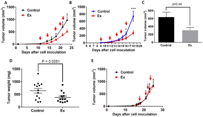
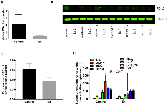
Biofield therapies have gained popularity and are being explored as possible treatments for cancer. In some cases, devices have been developed that mimic the electromagnetic fields that are emitted from people delivering biofield therapies. However, there is limited research examining if humans could potentially inhibit the proliferation of cancer cells and suppress tumor growth through modification of inflammation and the immune system. We found that human NSCLC A549 lung cancer cells exposed to Sean L. Harribance, a purported healer, showed reduced viability and downregulation of pAkt. We further observed that the experimental exposure slowed growth of mouse Lewis lung carcinoma evidenced by significantly smaller tumor volume in the experimental mice (274.3 ± 188.9 mm3) than that of control mice (740.5 ± 460.2 mm3; P < .05). Exposure to the experimental condition markedly reduced tumoral expression of pS6, a cytosolic marker of cell proliferation, by 45% compared with that of the control group. Results of reversed phase proteomic array suggested that the experimental exposure downregulated the PD-L1 expression in the tumor tissues. Similarly, the serum levels of cytokines, especially MCP-1, were significantly reduced in the experimental group ( P < .05). Furthermore, TILs profiling showed that CD8+/CD4- immune cell population was increased by almost 2-fold in the experimental condition whereas the number of intratumoral CD25+/CD4+ (T-reg cells) and CD68+ macrophages were 84% and 33%, respectively, lower than that of the control group. Together, these findings suggest that exposure to purported biofields from a human is capable of suppressing tumor growth, which might be in part mediated through modification of the tumor microenvironment, immune function, and anti-inflammatory activity in our mouse lung tumor model.
Keywords: Lewis lung carcinoma; PD-L1; biofield; immune modulation.
Conflict of interest statement
Figures


Similar articles
-
Human Biofield Therapy Modulates Tumor Microenvironment and Cancer Stemness in Mouse Lung Carcinoma.Integr Cancer Ther. 2020 Jan-Dec;19:1534735420940398. doi: 10.1177/1534735420940398. Integr Cancer Ther. 2020. PMID: 32975128 Free PMC article.
-
Stromal PD-L1-Positive Regulatory T cells and PD-1-Positive CD8-Positive T cells Define the Response of Different Subsets of Non-Small Cell Lung Cancer to PD-1/PD-L1 Blockade Immunotherapy.J Thorac Oncol. 2018 Apr;13(4):521-532. doi: 10.1016/j.jtho.2017.11.132. Epub 2017 Dec 18. J Thorac Oncol. 2018. PMID: 29269008
-
Bu Fei Decoction attenuates the tumor associated macrophage stimulated proliferation, migration, invasion and immunosuppression of non-small cell lung cancer, partially via IL-10 and PD-L1 regulation.Int J Oncol. 2017 Jul;51(1):25-38. doi: 10.3892/ijo.2017.4014. Epub 2017 May 19. Int J Oncol. 2017. PMID: 28534943 Free PMC article.
-
The lung microenvironment: an important regulator of tumour growth and metastasis.Nat Rev Cancer. 2019 Jan;19(1):9-31. doi: 10.1038/s41568-018-0081-9. Nat Rev Cancer. 2019. PMID: 30532012 Free PMC article. Review.
-
Biofield Physiology: A Framework for an Emerging Discipline.Glob Adv Health Med. 2015 Nov;4(Suppl):35-41. doi: 10.7453/gahmj.2015.015.suppl. Epub 2015 Nov 1. Glob Adv Health Med. 2015. PMID: 26665040 Free PMC article. Review.
Cited by
-
Recent Progress in Mind-Body Therapies in Cancer Care.Curr Oncol Rep. 2023 Apr;25(4):293-307. doi: 10.1007/s11912-023-01373-w. Epub 2023 Feb 8. Curr Oncol Rep. 2023. PMID: 36753025 Review.
-
Examining the effects of biofield therapy through simultaneous assessment of electrophysiological and cellular outcomes.Sci Rep. 2024 Dec 2;14(1):29221. doi: 10.1038/s41598-024-79617-3. Sci Rep. 2024. PMID: 39622875 Free PMC article.
-
Human Biofield Therapy Modulates Tumor Microenvironment and Cancer Stemness in Mouse Lung Carcinoma.Integr Cancer Ther. 2020 Jan-Dec;19:1534735420940398. doi: 10.1177/1534735420940398. Integr Cancer Ther. 2020. PMID: 32975128 Free PMC article.
-
Evaluation of the protective effect of Compound Kushen Injection against radiation‑induced lung injury in mice.Mol Med Rep. 2025 Apr;31(4):88. doi: 10.3892/mmr.2025.13453. Epub 2025 Feb 7. Mol Med Rep. 2025. PMID: 39917996 Free PMC article.
-
Differential In Vivo Effects on Cancer Models by Recorded Magnetic Signals Derived From a Healing Technique.Dose Response. 2023 Jun 2;21(2):15593258231179903. doi: 10.1177/15593258231179903. eCollection 2023 Apr-Jun. Dose Response. 2023. PMID: 37325440 Free PMC article.
References
-
- Hammerschlag R, Jain S, Baldwin AL, et al. Biofield research: a roundtable discussion of scientific and methodological issues. J Altern Complement Med. 2012;18:1081-1086. - PubMed
-
- Cohen L, Chen Z, Arun B, et al. External qigong therapy for women with breast cancer prior to surgery. Integr Cancer Ther. 2010;9:348-353. - PubMed
-
- Yan X, Shen H, Jiang H, Hu D, Wang J, Wu X. External Qi of Yan Xin Qigong inhibits activation of Akt, Erk1/2 and NF-kB and induces cell cycle arrest and apoptosis in colorectal cancer cells. Cell Physiol Biochem. 2013;31:113-122. - PubMed
Publication types
MeSH terms
Substances
Grants and funding
LinkOut - more resources
Full Text Sources
Medical
Research Materials
Miscellaneous
