

Anterolateral ligament of the knee: a step-by-step dissection
- PMID: 30947710
- PMCID: PMC6449941
- DOI: 10.1186/s12891-019-2517-0
Anterolateral ligament of the knee: a step-by-step dissection
Abstract
Background: The number of studies and clinical interest in the anterolateral ligament of the knee (ALL) has grown in recent years. A meticulous and accurate ALL dissection is vital in anatomic and biomechanical studies, and a standardized technique is not yet established. As such, the aim of this study was to describe a step-by-step ALL dissection technique that could help authors consistently identify the ALL.
Methods: Twenty knees from frozen adult cadavers, with no preference for sex or age, were included in the study. All the cadavers were dissected using the same technique to determine the incidence of the ALL.
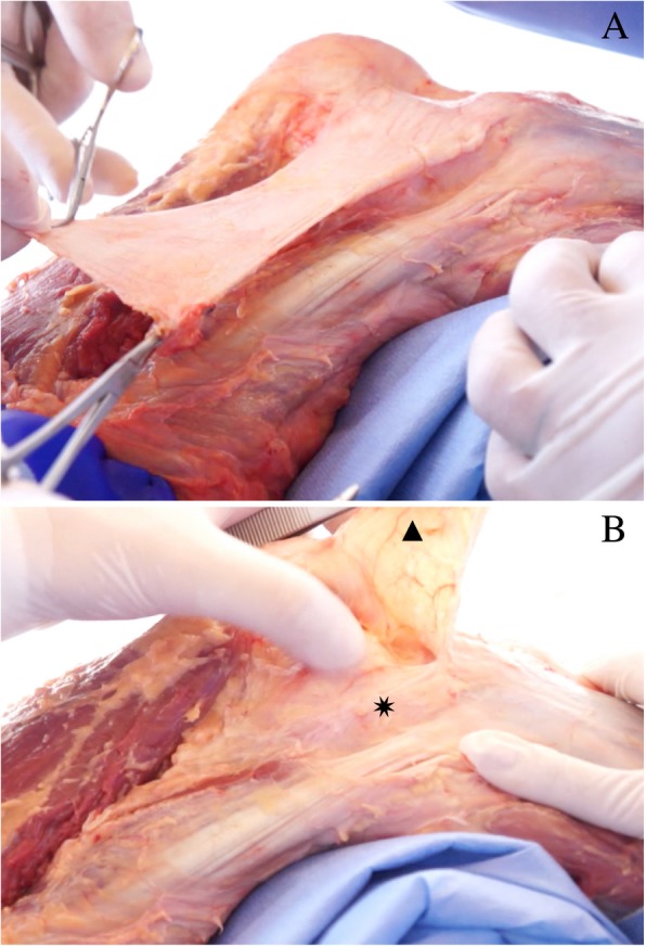
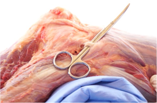
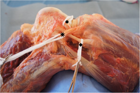
Results: A transverse incision is performed in the iliotibial band (ITB), around 10 cm proximal to the topography of the lateral epicondyle of the femur. Next, the ITB undergoes anterograde blunt dissection until its insertion at Gerdy's tubercle in the tibia. Maintaining biceps femoris insertion, a dissection is performed anteriorly to it, until the lateral collateral ligament (LCL) is found. Using the LCL, internal rotation and 30 to 60° flexion as references, the ALL can be located in the anterolateral topography of the knee, with its origin near the lateral epicondyle (proximal and posterior) and insertion between Gerdy's tubercle and the fibula (4.0 mm to 7.0 mm below the tibial plateau), expanding to the lateral meniscus (between the body and anterior horn), exhibiting a mean length of 4.0 ± 0.4 cm and mean width of 5.5 ± 0.8 mm.
Conclusions: The present article describes an effective and reproducible ALL dissection technique that made it was possible to identify the ligament in 100% of the cases in the present study.
Conflict of interest statement
Ethics approval and consent to participate
The study was approved by the Research Ethics Committee – Plataforma Brasil (CAAE: 78798617.5.0000.5049 - Brazil) and involved 20 knees from frozen adult cadavers, with no preference for sex or age. The cadavers were obtained from the Center for Forensic Studies of Ceará, Brazil (PEFOCE). The samples came from unclaimed cadavers after 30 days without contact from any relative or known, according to Local Law N° 8.501 of November 30, 1992.
Consent for publication
Not applicable.
Competing interests
The authors declare that they have no competing interests.
Publisher’s Note
Springer Nature remains neutral with regard to jurisdictional claims in published maps and institutional affiliations.
Figures


References
MeSH terms
LinkOut - more resources
Full Text Sources
Research Materials
