. 2019 Apr 4;16(1):10.
doi: 10.1186/s12987-019-0130-0.

Spinal CSF flow in response to forced thoracic and abdominal respiration

Affiliations

Spinal CSF flow in response to forced thoracic and abdominal respiration

Gökmen Aktas et al. Fluids Barriers CNS. .

Abstract

Background: Respiration-induced pressure changes represent a powerful driving force of CSF dynamics as previously demonstrated using flow-sensitive real-time magnetic resonance imaging (MRI). The purpose of the present study was to elucidate the sensitivity of CSF flow along the spinal canal to forced thoracic versus abdominal respiration.

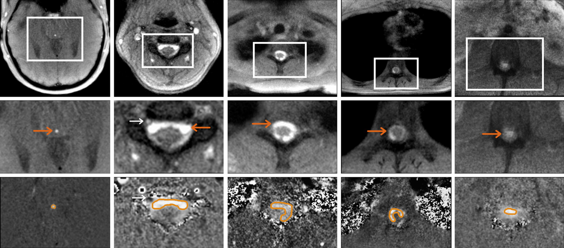
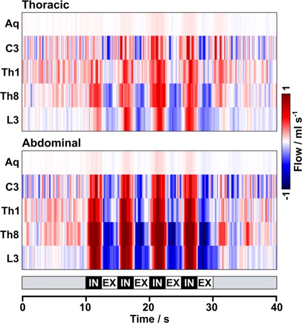
Methods: Eighteen subjects without known illness were studied using real-time phase-contrast flow MRI at 3 T in the aqueduct and along the spinal canal at levels C3, Th1, Th8 and L3. Subjects performed a protocol of forced breathing comprising four cycles of 2.5 s inspiration and 2.5 s expiration.

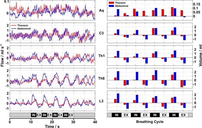
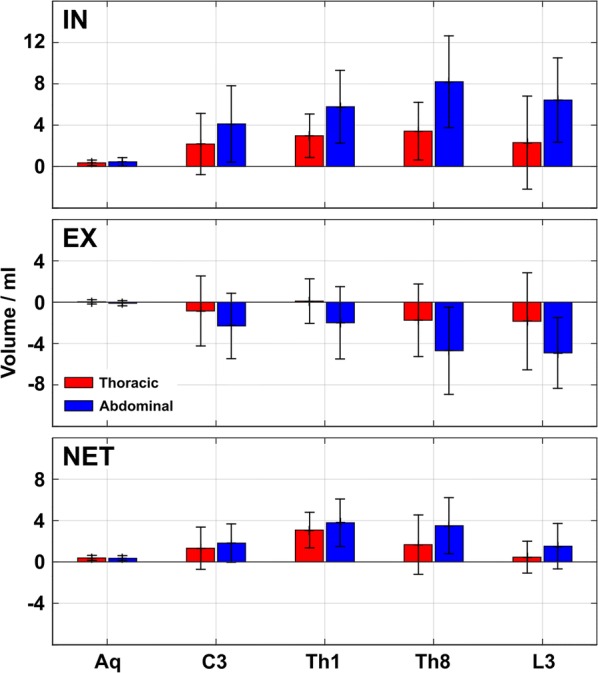
Results: The quantitative results for spinal CSF flow rates and volumes confirm previous findings of an upward movement during forced inspiration and reversed downward flow during subsequent exhalation-for both breathing types. However, the effects were more pronounced for abdominal than for thoracic breathing, in particular at spinal levels Th8 and L3. In general, CSF net flow volumes were very similar for both breathing conditions pointing upwards in all locations.

Conclusions: Spinal CSF dynamics are sensitive to varying respiratory performances. The different CSF flow volumes in response to deep thoracic versus abdominal breathing reflect instantaneous adjustments of intrathoracic and intraabdominal pressure, respectively. Real-time MRI access to CSF flow in response to defined respiration patterns will be of clinical importance for patients with disturbed CSF circulation like hydrocephalus, pseudotumor cerebri and others.

Keywords: CSF dynamics; Flow-sensitive real-time MRI; Hydrocephalus; Intraabdominal pressure; Intrathoracic pressure; Respiration.


Conflict of interest statement

The authors declare that they have no competing interests.

Figures

Fig. 1
Fig. 1
Regions of interest for CSF flow analyses. Top: magnitude images and Middle: magnified sections reveal CSF flow (bright signals, orange arrows ) during forced inspiration in (left to right) the aqueduct as well as at spinal level C3, Th1, Th8 and L3 of one representative subject (#7). Bottom: corresponding magnified velocity maps indicate upward flow during forced inspiration (bright signals, orange ROI), whereas epidural veins at spinal level C3 (white arrows) show concomitant downward flow of venous blood out of the head
Fig. 2
Fig. 2
Real-time MRI of thoracic and abdominal breathing. Selected sagittal real-time images through the right dome of the diaphragm during thoracic and abdominal respiration. Upper left: thoracic breathing at deep inspiration and upper right: deep expiration. Lower left: abdominal breathing at deep inspiration and lower right: deep expiration. Inspiration causes elevation of ribcage, widening of anteroposterior thoracic diameters, contraction of the diaphragm with downward movement of its dome and enlargement of the intrathoracic volume. The corresponding widening of the costodiaphragmatic recess (arrows) is more pronounced during abdominal inspiration
Fig. 3
Fig. 3
Mean CSF flow rates (ml s−1) during forced respiration. Mean color-coded flow rates averaged across subjects in the aqueduct as well as at spinal levels C3, Th1, Th8 and L3 for Top: forced thoracic and Bottom: forced abdominal breathing. The color scale was chosen to visualize the low flow in the aqueduct. Onset of forced inspiration prompts an increase of CSF flow in upward direction (red) at all locations, while downward movement (blue) prevails during expiration at all spinal levels. Forced abdominal breathing consistently causes higher flow rates compared to thoracic breathing
Fig. 4
Fig. 4
CSF dynamics during thoracic and abdominal breathing. Left: CSF flow in aqueduct and spinal canal (subject #16) during (red) thoracic or (blue) abdominal breathing. Right: CSF volumes during four cycles of forced respiration. The onset of forced inspiration leads to a distinct increase of upward CSF flow which is reversed during expiration. In spinal canal, abdominal breathing causes higher flow rates and volumes, which is not seen in the aqueduct (small flow rates and volumes). Aq aqueduct, C3 cervical level 3, Th1/Th8 thoracic levels 1 and 8, L3 lumbar level 3, IN inspiration, EX expiration
Fig. 5
Fig. 5
CSF net volumes during forced respiration. Mean CSF flow volumes averaged across 18 subjects and 4 cycles of forced thoracic (red) and abdominal (blue) Top: inspiration and Middle: expiration. Positive inspiratory CSF flow volumes indicate upward fluid movement which was more pronounced during abdominal breathing. Negative downward flow volumes during forced expiration varied, but showed a more uniform behavior during abdominal respiration. The large standard deviations during forced thoracic expiration indicate pronounced inter-individual differences. Bottom: CSF net volumes point upwards at all locations, again more distinct during abdominal breathing. IN inspiration, EX expiration, NET CSF net volumes

References

    1. Greitz D, Franck A, Nordell B. On the pulsatile nature of intracranial and spinal CSF-circulation demonstrated by MR imaging. Acta Radiol. 1993;34:321–328. doi: 10.1177/028418519303400403. - DOI - PubMed
    1. Enzmann DR, Pelc NJ. Normal flow patterns of intracranial and spinal cerebrospinal fluid defined with phase-contrast cine MR imaging. Radiology. 1991;178:467–474. doi: 10.1148/radiology.178.2.1987610. - DOI - PubMed
    1. Schroth G, Klose U. Cerebrospinal fluid flow. II. Physiology of respiration-related pulsations. Neuroradiology. 1992;35:10–15. doi: 10.1007/BF00588271. - DOI - PubMed
    1. Chen L, Beckett A, Verma A, Feinberg DA. Dynamics of respiratory and cardiac CSF motion revealed with real-time simultaneous multi-slice EPI velocity phase contrast imaging. Neuroimage. 2015;122:281–287. doi: 10.1016/j.neuroimage.2015.07.073. - DOI - PMC - PubMed
    1. Klose U, Strik C, Kiefer C, Grodd W. Detection of a relation between respiration and CSF pulsation with an echoplanar technique. J Magn Reson Imaging. 2000;11:438–444. doi: 10.1002/(SICI)1522-2586(200004)11:4<438::AID-JMRI12>3.0.CO;2-O. - DOI - PubMed

LinkOut - more resources