. 2019 Apr 4;38(1):145.
doi: 10.1186/s13046-019-1142-y.

Cx32 exerts anti-apoptotic and pro-tumor effects via the epidermal growth factor receptor pathway in hepatocellular carcinoma

Affiliations

Cx32 exerts anti-apoptotic and pro-tumor effects via the epidermal growth factor receptor pathway in hepatocellular carcinoma

Yuke Xiang et al. J Exp Clin Cancer Res. .

Abstract

Background: Abnormal expression or distribution of connexin 32 (Cx32) is associated with hepatocarcinogenesis, but the role of Cx32 and the underlying mechanisms are still unclear.

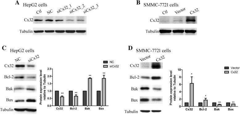
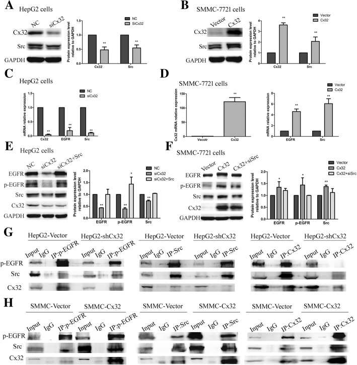
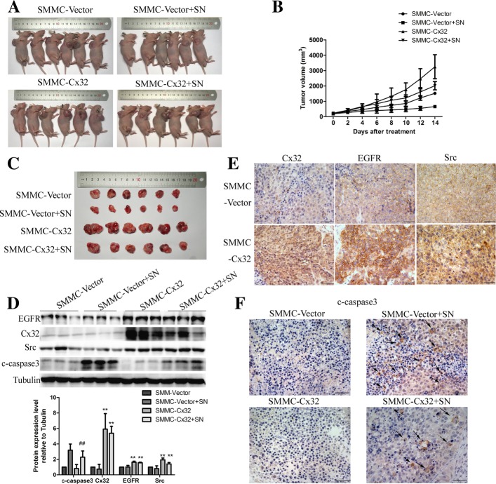
Methods: The expression level of Cx32 in 96 hepatocellular carcinoma (HCC) specimens was determined using western blotting and immunohistochemistry. The correlation between Cx32 expression and clinicopathological parameters was analyzed. The cell apoptosis rate was examined using flow cytometry and western blotting. The role of Cx32 in the Src kinase and epidermal growth factor receptor (EGFR) signaling pathways was measured by quantitative real-time PCR, western blotting and coimmunoprecipitation (CO-IP). The effect of Cx32 overexpression on the streptonigrin (SN)-induced tumor growth suppression and apoptosis was assessed in nude mice.

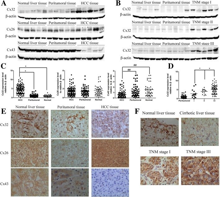
Results: Our study showed that overexpressed Cx32 accumulated in the cytoplasm and that Cx32-containing gap junctions (GJs) were nearly absent in HCC specimens. Upregulated Cx32 expression was highly correlated with advanced tumor-node-metastasis (TNM) stage and poor tumor differentiation and was an independent predictive marker for poor prognosis in HCC. Overexpression of Cx32 significantly inhibited SN-induced apoptosis by activating the EGFR signaling pathway in vitro and in vivo. Moreover, the expression levels of Cx32 and EGFR were positively correlated in HCC specimens. The CO-IP experiments demonstrated that Cx32 could bind to Src kinase, and the western blotting results revealed that Cx32 increased the levels of EGFR and p-EGFR by upregulating Src expression.

Conclusion: The present study demonstrated that overexpressed and internalized Cx32 was associated with advanced TNM stage and poor tumor differentiation and predicted poor prognosis in HCC. Cx32 facilitated HCC progression by blocking chemotherapy-induced apoptosis in vitro and in vivo via interacting with Src and thus promoting the phosphorylation of EGFR, subsequently activating the EGFR signaling pathway. Cx32 may be a potential biomarker and a new therapeutic target for HCC.

Keywords: Apoptosis; Connexin32; EGFR; Hepatocellular carcinoma; Nonjunctional function; Src.


Conflict of interest statement

Ethics approval and consent to participate

Ethical approval (K-201354)) of clinical data and tissues was obtained from the Research Committee of Ethics in the Affiliated Cancer Hospital of Xinjiang Medical University. Written informed consent form for the experimental studies was obtained from the patients or their guardians. All the animal experimental procedures were approved by the Institutional Animal Care and Use Committee of Sun Yat-sen University.

Consent for publication

All authors agree for publication.

Competing interests

All authors read and approved the final version of the manuscript, and the authors declare that they have no competing interests.

Publisher’s Note

Springer Nature remains neutral with regard to jurisdictional claims in published maps and institutional affiliations.

Figures

Fig. 1
Fig. 1
Expression and distribution of Cx32, Cx26 and Cx43 in patients with HCC. a. The protein expression levels of Cx32, Cx26 and Cx43 were determined by western blot analysis. β-actin was used as the loading control. b. The expression of Cx32 was correlated with increased TNM stages, as revealed by western blot analysis. β-actin was used as the loading control. c. Statistical analysis of the relative expression levels of Cxs in HCC tissues, peritumoral tissues, and normal liver tissues. **, P < 0.01; ##, P < 0.01. d. Statistical analysis of the relative expression levels of Cx32 in peritumoral tissues and HCC tissues with different TNM stages. *, P < 0.05. e. Representative IHC staining of Cx32, Cx26 and Cx43 protein in normal liver tissues (left panels), peritumoral tissues (middle panels) and HCC tissues (right panels) (400×). Scale bars: 50 μm. f. Representative IHC staining of Cx32 in normal liver tissues, cirrhotic tissues and early and advanced HCC tissues (400×). Scale bars: 50 μm
Fig. 2
Fig. 2
Kaplan-Meier analysis. Patients in the low Cx32 group (n = 48) had significantly longer overall survival (OS) times than those in the high Cx32 group (n = 48) (P = 0.014, log-rank test)
Fig. 3
Fig. 3
Cx32 regulates the expression of Bcl-2 family proteins in HCC cell lines. a. Cx32 expression was knocked down in HepG2 cells by siRNA transfection. siCx32_2 showed the greatest efficiency in reducing Cx32 expression. b. Transient plasmid transfection into SMMC-7721 cells induced Cx32 overexpression. c. Silencing Cx32 expression in HepG2 cells increased the expression levels of Bax and Bak but decreased the expression level of Bcl-2 (n = 3). **, P < 0.01 versus NC. d. Overexpression of Cx32 in SMMC-7721 cells caused the upregulation of Bcl-2 expression and the downregulation of Bax and Bak expression (n = 3). **, P < 0.01 versus Vector
Fig. 4
Fig. 4
Cx32 exerts an anti-apoptotic effect on HepG2 cells in a GJ-independent manner. a. When GJ function was inhibited by pretreatment with 2-APB (50 μm, 2 h), knockdown of Cx32 promoted the SN-induced increase in the levels of cleaved-caspase3 and cleaved-PRAR in HepG2 cells, as revealed by western blot analysis (n = 3). **, P < 0.01; ##, P < 0.01. b. Silencing the expression of Cx32 promoted SN-induced apoptosis in HepG2 cells when GJ function was inhibited by 2-APB, as assessed by flow cytometry (n = 3). ##, P < 0.01 versus NC; **, P < 0.01 versus NC + 2-APB + SN. c. When GJs were physically eliminated by low-density culture conditions, Cx32 silencing enhanced the SN-induced increase in the levels of cleaved-caspase3 and cleaved-PARP in HepG2 cells, as revealed by western blot analysis (n = 3). **, P < 0.01; ##, P < 0.01. d. Cx32 silencing facilitated SN-induced apoptosis in HepG2 cells in low-density culture, as assessed by flow cytometry (n = 3). **, P < 0.01 versus NC + SN
Fig. 5
Fig. 5
Overexpressed Cx32 exerts an anti-apoptotic effect on SMMC-7721 cells in a GJ-independent manner. a. When GJ function was inhibited by pretreatment with 2-APB, overexpression of Cx32 alleviated the SN-induced increase in the levels of cleaved-caspase3 and cleaved-PARP in SMMC-7721 cells, as revealed by western blot analysis (n = 3). **, P < 0.01; ##, P < 0.01. b. Upregulation of Cx32 expression suppressed SN-induced apoptosis in SMMC-7721 cells when GJ function was inhibited by 2-APB, as revealed by flow cytometry (n = 3). **, P < 0.01 versus Cx32 + 2-APB + SN. c. When GJ were physically eliminated by low-density culture conditions, Cx32 overexpression reduced SN-induced increase in the levels of cleaved-caspase3 and cleaved-PARP in SMMC-7721 cells (n = 3). **, P < 0.01; ##, P < 0.01. d. Upregulation of Cx32 expression inhibited SN-induced apoptosis in SMMC-7721 cells in low-density culture, as revealed by flow cytometry (n = 3). **, P < 0.01 versus Cx32 + SN
Fig. 6
Fig. 6
Cx32 exerts anti-apoptotic effects by activating EGFR signaling pathway. a. In 30 HCC specimens, the expression of EGFR was significantly correlated with the expression of Cx32 (r = 0.662, P < 0.01). b. The expression level of EGFR was significantly higher in HepG2 cells than in SMMC-7721 cells. c and d. The effects of Cx32 knockdown or overexpression on the EGFR signaling pathway in HepG2 cells and SMMC-7721 cells were determined by western blot analysis (n = 3, respectively). **, P < 0.01 versus HepG2 NC (c) or SMMC-7721 Vector (d). e. In rescue experiments, cotransfection of siRNA-Cx32 and EGFR-expression vectors into HepG2 cells reversed the pro-apoptotic effects of Cx32 knockdown. **, P < 0.01; ##, P < 0.01. f. In rescue experiments, cotransfection of Cx32 expression vectors and siRNA-EGFR into SMMC-7721 cells reversed the anti-apoptotic effects of Cx32 overexpression. **, P < 0.01; ##, P < 0.01
Fig. 7
Fig. 7
Cx32 upregulates the expression and activation of EGFR by binding with Src. a and b. In HCC cell lines, silencing or overexpressing Cx32 caused Src downregulation or upregulation, respectively. **, P < 0.01 versus HepG2 NC (a) or SMMC-7721 Vector (b). c. Silencing Cx32 by siCx32 transfection caused a decrease in the EGFR and Src mRNA levels in HepG2 cells, as determined by qPCR. GAPDH was used as the loading control. **, P < 0.01 versus NC. d. Overexpression of Cx32 in SMMC-7721 cells upregulated the mRNA levels of Cx32 (left panel), EGFR and Src (right panel), as determined by qPCR. GAPDH was used as the loading control. **, P < 0.01 versus Vector. e. The decrease in the levels of EGFR, p-EGFR and Src mediated by siCx32 was reversed by cotransfection of the Src overexpression vector in HepG2 cells. **, P < 0.01 versus NC. f. The increase in the levels of EGFR, p-EGFR and Src induced by Cx32 overexpression was rescued by cotransfection of siSrc in SMMC-7721 cells. **, P < 0.01 versus Vector. g and h. Cx32, p-EGFR and Src interacted with each other, as detected by CO-IP experiments in HepG2 cells (g) and SMMC-7721 cells (h). Lysate supernatants incubated without antibody, termed Input, were used as the positive control, and proteins precipitated by IgG were used as the negative control
Fig. 8
Fig. 8
Overexpression of Cx32 promotes the proliferation of SMMC-7721 cells and protects cells from SN-induced apoptosis in vivo. a. Representative images of the nude mouse xenograft model. b. Tumor growth curves. Overexpression of Cx32 promoted the tumor growth in nude mice and significantly reduced the growth suppression mediated by intragastric injection of SN (0.5 mg/kg). c. Representative images of tumors from the sacrificed nude mice. d. Representative images of IHC for Cx32, EGFR and Src in tumors generated from SMMC-Vector and SMMC-Cx32 cells. Scale bars: 50 μm. e. Overexpression of Cx32 inhibited the SN-induced increase in the levels of cleaved-caspase3 by increasing the levels of EGFR and Src. ##, P < 0.01 vs Vector +SN. **, P < 0.01 versus Vector. f. Representative images of IHC for cleaved-caspase3 in each group. Overexpression of Cx32 inhibited the SN-induced increase in the level of cleaved-caspase3. Scale bars: 50 μm

References

    1. Beyer EC, Berthoud VM. Gap junction gene and protein families: Connexins, innexins, and pannexins. Biochim Biophys Acta Biomembr. 2018;1860:5–8. doi: 10.1016/j.bbamem.2017.05.016. - DOI - PMC - PubMed
    1. Beyer EC, Berthoud VM. Gap junction structure: unraveled, but not fully revealed. F1000Research. 2017;6:568. doi: 10.12688/f1000research.10490.1. - DOI - PMC - PubMed
    1. Aasen T, Mesnil M, Naus CC, Lampe PD, Laird DW. Gap junctions and cancer: communicating for 50 years. Nat Rev Cancer. 2016;16:775–788. doi: 10.1038/nrc.2016.105. - DOI - PMC - PubMed
    1. Mao XY, Li QQ, Gao YF, Zhou HH, Liu ZQ, Jin WL. Gap junction as an intercellular glue: emerging roles in cancer EMT and metastasis. Cancer Lett. 2016;381:133–137. doi: 10.1016/j.canlet.2016.07.037. - DOI - PubMed
    1. Zhang Y, Tao L, Fan L, Peng Y, Yang K, Zhao Y, et al. Different gap junction-propagated effects on cisplatin transfer result in opposite responses to cisplatin in normal cells versus tumor cells. Sci Rep. 2015;5:12563. doi: 10.1038/srep12563. - DOI - PMC - PubMed