. 2019 Mar:115:14-23.
doi: 10.1016/j.tube.2019.01.002. Epub 2019 Jan 16.

Mycobacteriophage ZoeJ: A broad host-range close relative of mycobacteriophage TM4

Affiliations

Mycobacteriophage ZoeJ: A broad host-range close relative of mycobacteriophage TM4

Rebekah M Dedrick et al. Tuberculosis (Edinb). 2019 Mar.

Abstract

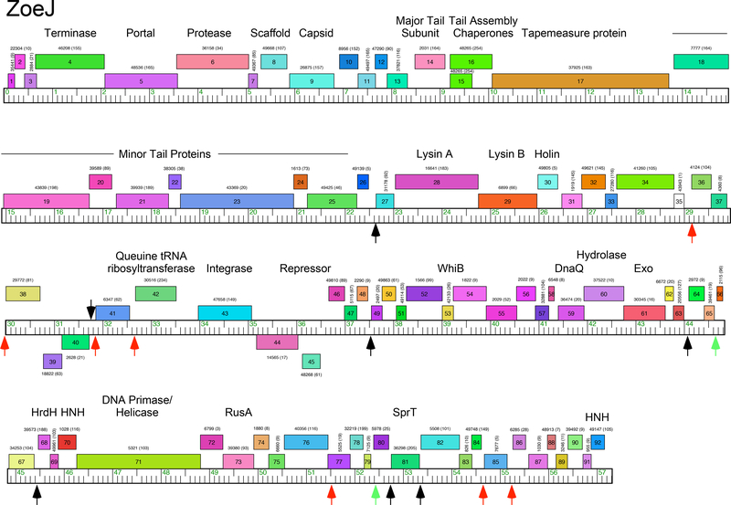
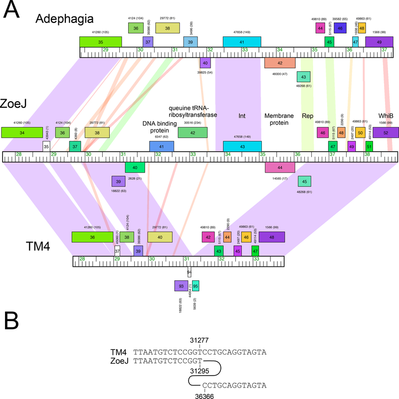
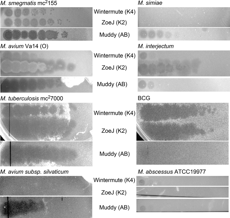
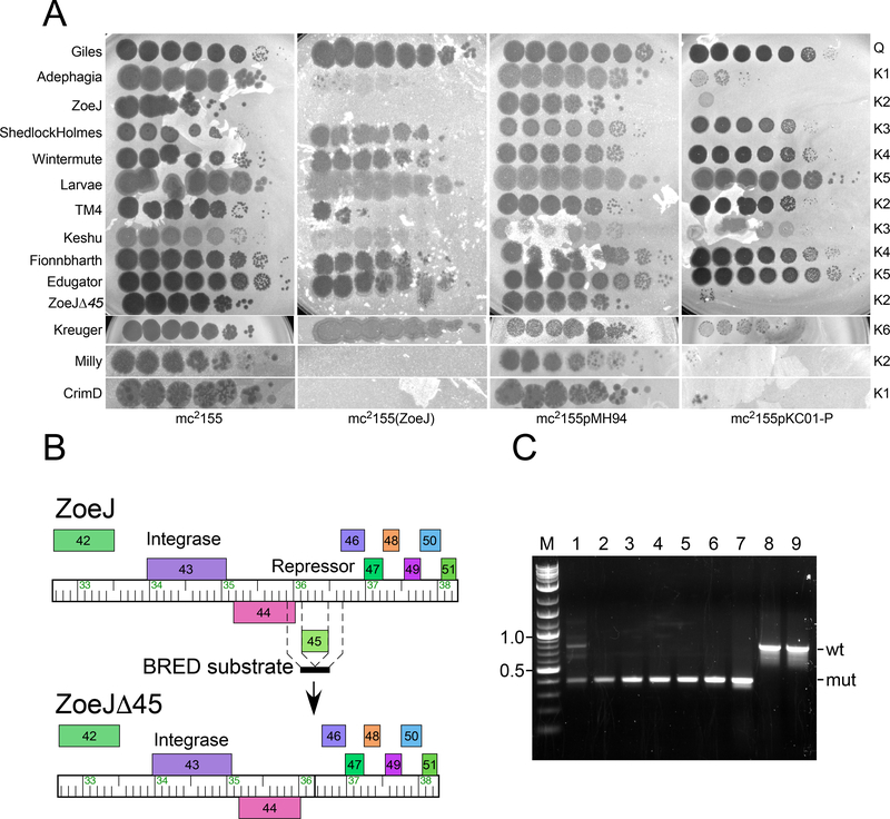
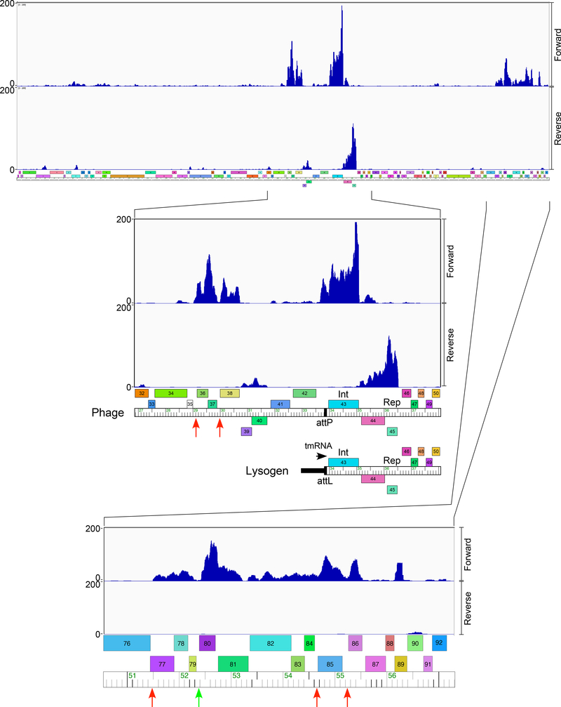
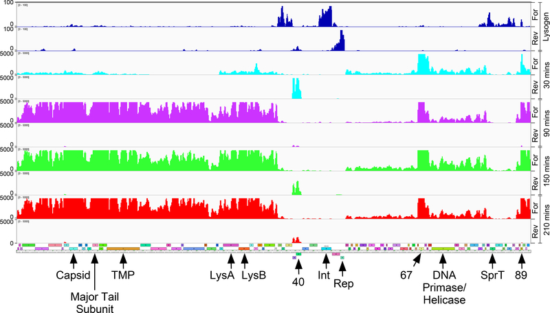
A collection of over 1600 sequenced bacteriophages isolated on a single host strain, Mycobacterium smegmatis mc2155, can be grouped into over two dozen types that have little or no nucleotide sequence similarity to each other. One group, Cluster K, can be divided into several subclusters, and the well-characterized and much exploited phage TM4 lies in Subcluster K2. Many of the Cluster K phages have broad host ranges and infect both fast- and slow-growing mycobacterial strains. Here we describe phage ZoeJ, a new Subcluster K2 member, which infects a broad spectrum of mycobacterial hosts including M. smegmatis, Mycobacterium tuberculosis, and Mycobacterium avium. ZoeJ has extensive sequence similarity to TM4, and comparative analysis reveals the precise deletion conferring the lytic phenotype of TM4. The ZoeJ immunity repressor was identified as gene 45, which is prophage-expressed, is required for lysogeny, and is sufficient to confer superinfection immunity to ZoeJ. ZoeJ gp45 also confers immunity to Subcluster K2 phage Milly, and Subcluster K1 phages Adephagia and CrimD, but surprisingly not to TM4. RNAseq analysis reveals the temporal pattern of early and late gene expressions in ZoeJ lytic growth and suggests a role for the ESAS motifs for gene regulation.


Figures

Figure 1.
Figure 1.. Genome map of mycobacteriophage ZoeJ.
The genome of ZoeJ is represented as a ruler with kbp markers with the predicted genes as colored boxes above or below the genome corresponding to rightwards- and leftwards-transcription, respectively. Gene names are shown within each box, and the corresponding phamily number shown above or below each gene with the number of phamily members in parentheses. The map was drawn using Phamerator and the database ‘actinobacteriophages_draft’ as of July 2018. Black and red vertical arrows indicate the locations of Start Associated Sequences (SAS; see Table 1), with the red arrows indicating those that also include an Extended Start Associated Sequence (ESAS; see Table 2). Green vertical arrows indicate positions of ESAS site that lack a linked SAS. Putative gene functions are indicated.
Figure 2.
Figure 2.. Mapping a deletion in the phage TM4 genome.
A. Genome maps of the central portions of phages Adephagia, ZoeJ, and TM4 are shown, with pairwise nucleotide sequence similarity shown as spectrum-colored shading between genomes; violet indicates closest similarity, and red the least similarity above a threshold E value of 10-4. Maps are annotated as in Fig. 1. B. Alignment of TM4 and ZoeJ shows that the deletion in TM4 occurred precisely between ZoeJ coordinates 31,295 and 36,366.
Figure 3.
Figure 3.. Host range of mycobacteriophage ZoeJ.
Lawns of mycobacterial strains were spotted with ten-fold serial dilutions (left to right) of Cluster K phages ZoeJ (Subcluster K2) and Wintermute (Subcluster K4) together with control phage Muddy (Cluster AB) and incubated. The highest titer phage spot corresponds to a 10−1 dilution of the stock lysate of each phage.
Figure 4.
Figure 4.. ZoeJ gene 45 codes for repressor-mediated superinfection immunity.
A. Lawns of four strains – M. smegmatis mc2155, a ZoeJ lysogen [mc2155(ZoeJ)], a vector-containing strain (mc2155pMH94), and a strain expressing the ZoeJ repressor (mc2155pKC01-P) were spotted with ten-fold serial dilutions of phages as indicated at the left. The highest titer phage spot corresponds to a 10−1 dilution of the stock lysate of each phage. The cluster designation is shown at the right. B. Scheme for construction of the ZoeJΔ45 mutant. The position of the 400 bp BRED dsDNA substrate is shown with 200 bp of sequence identities flanking gene 45. Recombination between phage genomic DNA and the BRED substrate results in deletion of gene 45. C. Identification of a ZoeJΔ45 mutant by PCR. Following co-electroporation of ZoeJ genomic DNA and the BRED substrates, primary plaques were screened by PCR, a mixed primary plaque (lane 1) contains both wild-type (wt; 770 bp) and mutant (mut; 400 bp) products was identified. After replating, an isolated plaque containing only the mutant allele (lane 3) was identified, and after purification, four individual plaques (lanes 4–7) all have only the mutant allele. Wild-type ZoeJ phage lysate (lane 8) and ZoeJ phage DNA (lane 9) contain only the wild type allele. M: DNA Marker, sizes shown in kbp.
Figure 5.
Figure 5.. ZoeJ lysogenic transcription.
Lysogenic expression of ZoeJ prophage genes is shown at the top, with expanded views of the att-proximal region and the extreme genome right end are shown below. RNAseq reads are mapped to the ZoeJ prophage, but are represented on the linear viral form of the genome. The expanded view of the central genome part includes a map indicating the prophage orientation near the attL integration junction. The RNAseq reads are strand-specific and those mapping to forward and reverse DNA orientations are indicated. Locations of ESAS motifs associated with either an SAS motif or not (red and green respectively) are shown as vertical arrows. We note that although the overall expression patterns are similar in duplicate experiments with independent lysogenic cultures that there are subtle variations in relative gene expression. Data for an independently tested lysogen are shown in Figure S3.
Figure 6.
Figure 6.. ZoeJ transcription in lytic growth.
RNA was isolated at 30, 90, 150, and 210 minutes (shown in aqua, purple, green, and red, respectively) after infection of M. smegmatis with ZoeJ; reads mapping to forward (For) and reverse (Rev) strands are shown as indicated. A map of the ZoeJ genome is shown below (see Fig. 1 for details). The lysogen RNAseq data is shown for comparison in blue at the top. The positions of several key genes are indicated (9, capsid subunit; 14, major tail subunit; 45, repressor; TMP, Tape Measure Protein).

References

    1. Hatfull GF. Molecular Genetics of Mycobacteriophages. Microbiology Spectrum 2014;2:1–36. doi: doi:10.1128/microbiolspec.MGM2-0032-2013 - DOI - PMC - PubMed
    1. Hatfull GF. Mycobacteriophages: windows into tuberculosis. PLoS Pathog 2014;10:e1003953. doi: 10.1371/journal.ppat.1003953 - DOI - PMC - PubMed
    1. Jacobs-Sera D, Marinelli LJ, Bowman C, Broussard GW, Guerrero Bustamante C, Boyle MM, Petrova ZO, Dedrick RM, Pope WH, Science Education Alliance Phage Hunters Advancing G, Evolutionary Science Sea-Phages P, Modlin RL, Hendrix RW, Hatfull GF. On the nature of mycobacteriophage diversity and host preference. Virology 2012;434:187–201. doi: 10.1016/j.virol.2012.09.026 - DOI - PMC - PubMed
    1. Russell DA, Hatfull GF. PhagesDB: the actinobacteriophage database. Bioinformatics 2017;33:784–786. doi: 10.1093/bioinformatics/btw711 - DOI - PMC - PubMed
    1. Pope WH, Mavrich TN, Garlena RA, Guerrero-Bustamante CA, Jacobs-Sera D, Montgomery MT, Russell DA, Warner MH, Science Education Alliance-Phage Hunters Advancing G, Evolutionary S, Hatfull GF. Bacteriophages of Gordonia spp. Display a Spectrum of Diversity and Genetic Relationships. MBio 2017;8 doi: 10.1128/mBio.01069-17 - DOI - PMC - PubMed

Publication types

MeSH terms

LinkOut - more resources