

Non-Contrast-Enhancing Tumor: A New Frontier in Glioblastoma Research
- PMID: 30948373
- PMCID: PMC7053910
- DOI: 10.3174/ajnr.A6025
Non-Contrast-Enhancing Tumor: A New Frontier in Glioblastoma Research
Abstract
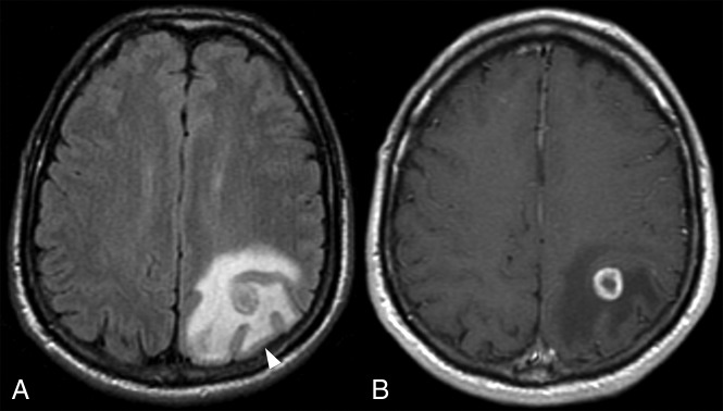
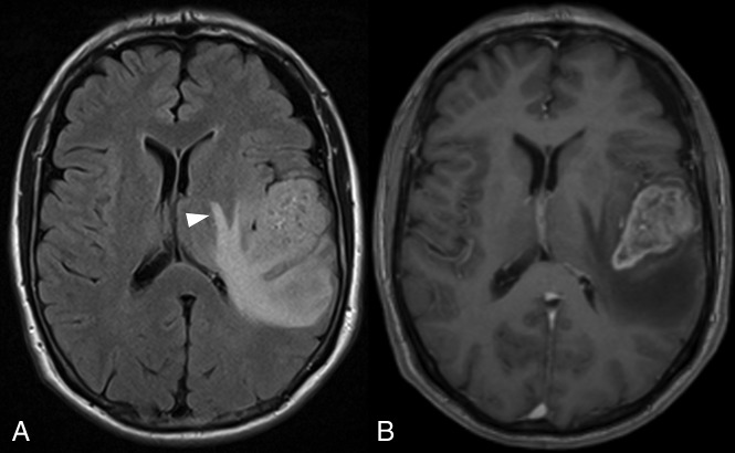
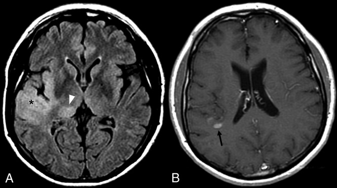
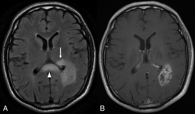
There is a growing understanding of the prognostic importance of non-contrast-enhancing tumor in glioblastoma, and recent attempts at more aggressive management of this component using neurosurgical resection and radiosurgery have been shown to prolong survival. Optimizing these therapeutic strategies requires an understanding of the features that can distinguish non-contrast-enhancing tumor from other processes, in particular vasogenic edema; however, the limited and heterogeneous manner in which it has been defined in the literature limits clinical translation. This review covers pertinent literature on our growing understanding of non-contrast-enhancing tumor and focuses on key conventional MR imaging features for improving its delineation. Such features include subtle differences in the degree of FLAIR hyperintensity, gray matter involvement, and focal mass effect. Improved delineation of tumor from edema will facilitate more aggressive management of this component and potentially realize associated survival benefits.
© 2019 by American Journal of Neuroradiology.
Figures
References
-
- Louis DN, Ohgaki H, Wiestler OD, et al. . World Health Organization Histological Classification of Tumors of the Central Nervous System. Lyon: International Agency for Research on Cancer; 2016
-
- Weller M, van den Bent M, Tonn JC, et al. ; European Association for Neuro-Oncology (EANO) Task Force on Gliomas. European Association for Neuro-Oncology (EANO) guideline on the diagnosis and treatment of adult astrocytic and oligodendroglial gliomas. Lancet Oncol 2017;18:e315–29 10.1016/S1470-2045(17)30194-8 - DOI - PubMed
Publication types
MeSH terms
LinkOut - more resources
Full Text Sources
Medical