Review
. 2019 May;20(5):e47560.
doi: 10.15252/embr.201847560. Epub 2019 Apr 4.

Shieldin - the protector of DNA ends

Affiliations
Review

Shieldin - the protector of DNA ends

Dheva Setiaputra et al. EMBO Rep. 2019 May.

Abstract

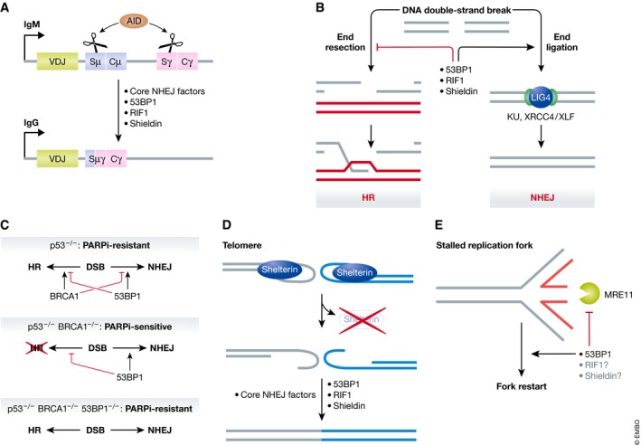
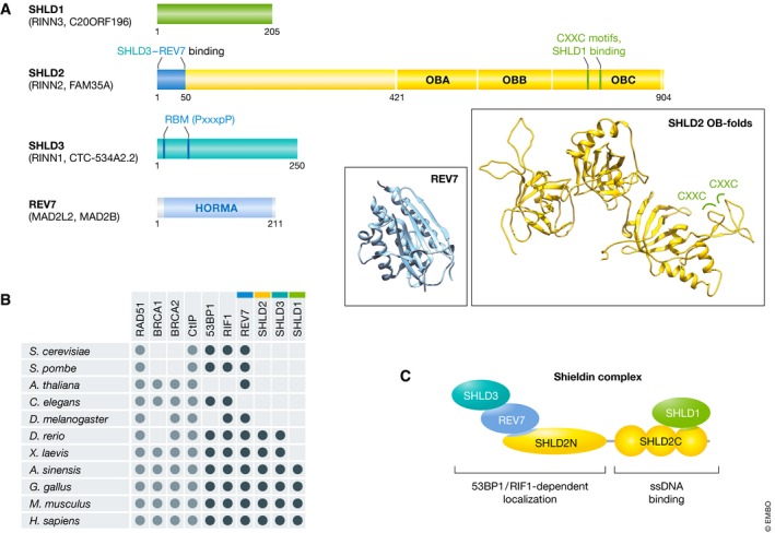
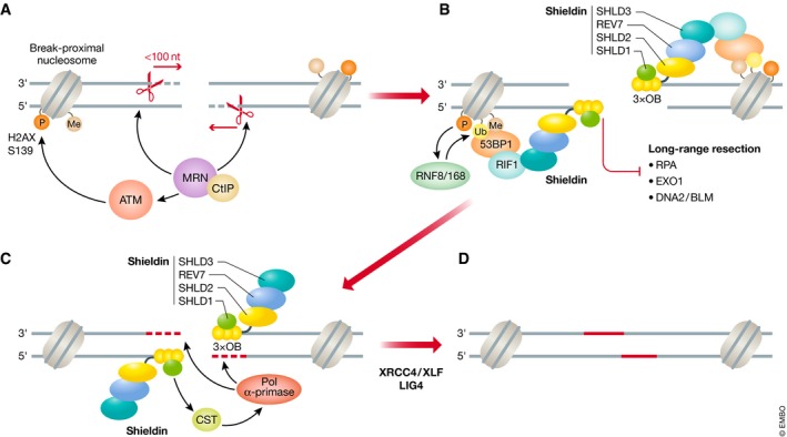
DNA double-strand breaks are a threat to genome integrity and cell viability. The nucleolytic processing of broken DNA ends plays a central role in dictating the repair processes that will mend these lesions. Usually, DNA end resection promotes repair by homologous recombination, whereas minimally processed ends are repaired by non-homologous end joining. Important in this process is the chromatin-binding protein 53BP1, which inhibits DNA end resection. How 53BP1 shields DNA ends from nucleases has been an enduring mystery. The recent discovery of shieldin, a four-subunit protein complex with single-stranded DNA-binding activity, illuminated a strong candidate for the ultimate effector of 53BP1-dependent end protection. Shieldin consists of REV7, a known 53BP1-pathway component, and three hitherto uncharacterized proteins: C20orf196 (SHLD1), FAM35A (SHLD2), and CTC-534A2.2 (SHLD3). Shieldin promotes many 53BP1-associated activities, such as the protection of DNA ends, non-homologous end joining, and immunoglobulin class switching. This review summarizes the identification of shieldin and the various models of shieldin action and highlights some outstanding questions requiring answers to gain a full molecular understanding of shieldin function.

Keywords: DNA repair; end resection; genome stability; homologous recombination; non‐homologous end joining.


Conflict of interest statement

DS has no conflict of interest to declare; DD declares that he is a founder and shareholder of Repare Therapeutics.

Figures

Figure 1
Figure 1. 53BP1 and shieldin act in various physiological contexts
(A) 53BP1, RIF1, and shieldin mediate immunoglobulin class switch recombination. During B‐cell stimulation, the activation‐induced cytidine deaminase (AID) enzyme causes single‐stranded breaks at two switch regions within the immunoglobulin heavy chain locus. 53BP1, RIF1, and shieldin are essential for non‐homologous end joining (NHEJ)‐mediated fusion of the two distant switch regions, altering the antibody subtype expressed from the locus. (B) Genomic DNA double‐strand breaks can be repaired through two competing pathways: the resection‐dependent homologous recombination or resection‐independent direct ligation through NHEJ. The components of the 53BP1 pathway inhibit end resection and facilitate repair via NHEJ. (C) 53BP1 and BRCA1 antagonize each other. In a Δp53 background, BRCA1 promotes HR and inhibits NHEJ while 53BP1 promotes NHEJ and inhibits HR, resulting in both DSB repair pathways being active. HR‐proficient cells are resistant to PARP inhibition (PARPi). In the absence of BRCA1, 53BP1 inhibits HR, resulting in HR deficiency and PARPi sensitivity. Concurrent depletion of BRCA1 and 53BP1 results in the de‐repression of HR, resulting in PARPi resistance. BRCA1 depletion is lethal in p53‐proficient cells unless accompanied by a depletion of 53BP1. (D) Telomere dysfunction due to shelterin subunit depletion results in aberrant DNA end processing. TRF2 depletion results in 53BP1‐dependent fusion of telomeres. (E) 53BP1 prevents MRE11‐mediated degradation of stalled replication forks. During DNA replication, stalled replication forks can reverse into the “chicken‐foot” configuration depicted. The nascent DNA portion (red) is a substrate for MRE11‐mediated exonucleolytic degradation. 53BP1 prevents this degradation and promotes fork restart, while the involvement of RIF1 and shieldin in this context has not been characterized.
Figure 2
Figure 2. Schematic of shieldin subunits and the architecture of the complex
(A) Amino acid residues of interest are numbered over each shieldin component. The three predicted tandem oligonucleotide/oligosaccharide‐binding (OB) folds in SHLD2 and the HORMA domain of REV7 are depicted. SHLD2 contains a CXXC zinc‐finger motif while SHLD3 has a REV7‐binding motif (RBM). Within the RBM (PxxxpP), P is an essential proline, p denotes an optional proline, and x represents any amino acid. Ribbon structures: The predicted structure of the SHLD2 OB‐folds (generated by homology modeling using the RPA1 structure; PDB:4GNX) with two CXXC zinc‐finger motifs highlighted is shown in yellow. The structure of REV7 (PDB:3ABE) is shown in cyan. (B) Evolutionary conservation of shieldin and other proteins involved in DNA double‐strand break repair pathway choice, based on known orthologues and BLAST homology search. (C) Functional architecture of the shieldin complex. SHLD3 and REV7 associate with the SHLD2 N‐terminus, forming the 53BP1‐ and RIF1‐dependent localization module. Meanwhile, SHLD1 associates with the SHLD2 C‐terminus, forming the ssDNA‐binding module.
Figure 3
Figure 3. Proposed 53BP1‐RIF1‐shieldin mechanism of action in DNA double‐strand break repair
(A) The MRE11‐RAD50‐NBS1 (MRN) complex and its accessory factor CtIP is recruited to DNA double‐strand break sites and introduces an endonuclease nick on the 5′‐terminated strand, which is expanded toward the break via the 3′–5′ exonuclease activity of MRE11. The nucleolytic activity of MRN results in short (< 100 nucleotides) tracts of single‐stranded (ss) DNA around the break. The NBS1 subunit of MRN also recruits and activates the ataxia telangiectasia‐mutated (ATM) kinase that phosphorylates histone H2AX at serine 139 (γH2A.X) on nucleosomes surrounding the DSB. (B) γH2AX recruits the RNF8 ubiquitin ligase through MDC1 binding (not shown). RNF8 catalyzes the K63‐linked polyubiquitination of histone H1, which in turn recruits RNF168. RNF168 ubiquitinates histone H2A lysine 15 which, in concert with constitutive H4 lysine 20 methylation, recruits 53BP1. 53BP1 recruits RIF1 in an ATM‐dependent manner, which localizes the shieldin complex to the DSB. The three tandem OB‐folds of SHLD2 binds the short ssDNA tract and inhibits long‐range 5′–3′ resection mediated by EXO1 and DNA2/BLM nucleases. (C) Shieldin then recruits the CTC1‐STN1‐TEN1 (CST) complex and its binding partner, the polymerase α‐primase (Pol α‐primase) complex to the DSB site. Pol α‐primase fills in the short ssDNA tract by synthesizing new DNA (red). (D) The DSB is subsequently repaired by non‐homologous end joining via DNA ligase IV (LIG4) and its accessory factors XRCC4/XLF. P, Ub, and Me represent phosphorylated, ubiquitinated, and methylated histones, respectively. S1 represents SHLD1. Black dashed lines indicate short range resection, while red dashed lines indicate fill‐in synthesis.

References

    1. Panier S, Boulton SJ (2014) Double‐strand break repair: 53BP1 comes into focus. Nat Rev Mol Cell Biol 15: 7–18 - PubMed
    1. Bunting SF, Callen E, Wong N, Chen HT, Polato F, Gunn A, Bothmer A, Feldhahn N, Fernandez‐Capetillo O, Cao L et al (2010) 53BP1 inhibits homologous recombination in Brca1‐deficient cells by blocking resection of DNA breaks. Cell 141: 243–254 - PMC - PubMed
    1. Nakamura K, Sakai W, Kawamoto T, Bree RT, Lowndes NF, Takeda S, Taniguchi Y (2006) Genetic dissection of vertebrate 53BP1: a major role in non‐homologous end joining of DNA double strand breaks. DNA Repair 5: 741–749 - PubMed
    1. Ward IM, Reina‐San‐Martin B, Olaru A, Minn K, Tamada K, Lau JS, Cascalho M, Chen L, Nussenzweig A, Livak F et al (2004) 53BP1 is required for class switch recombination. J Cell Biol 165: 459–464 - PMC - PubMed
    1. Manis JP, Morales JC, Xia Z, Kutok JL, Alt FW, Carpenter PB (2004) 53BP1 links DNA damage‐response pathways to immunoglobulin heavy chain class‐switch recombination. Nat Immunol 5: 481–487 - PubMed

Publication types

MeSH terms