. 2019 May 17;294(20):8184-8196.
doi: 10.1074/jbc.RA118.006920. Epub 2019 Apr 4.

Dephosphorylation of the transcriptional cofactor NACA by the PP1A phosphatase enhances cJUN transcriptional activity and osteoblast differentiation

Affiliations

Dephosphorylation of the transcriptional cofactor NACA by the PP1A phosphatase enhances cJUN transcriptional activity and osteoblast differentiation

William N Addison et al. J Biol Chem. .

Abstract

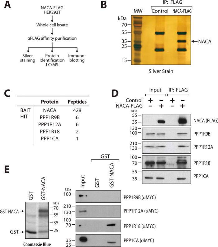
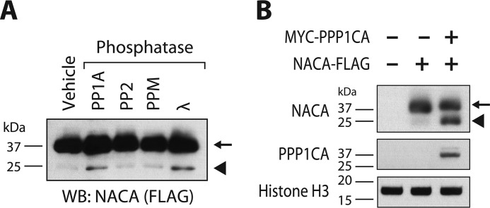
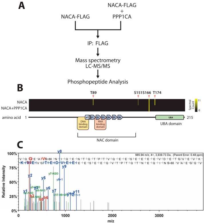
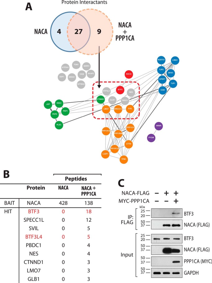
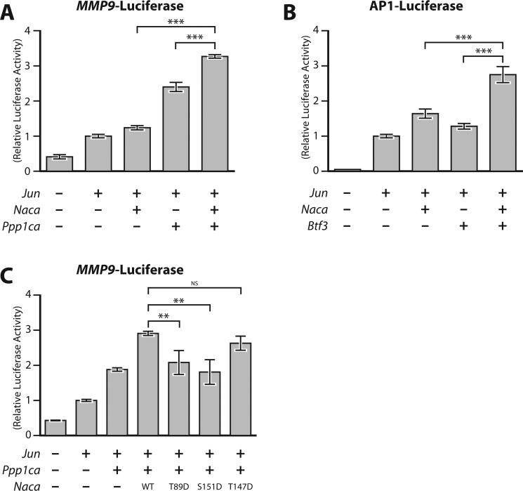
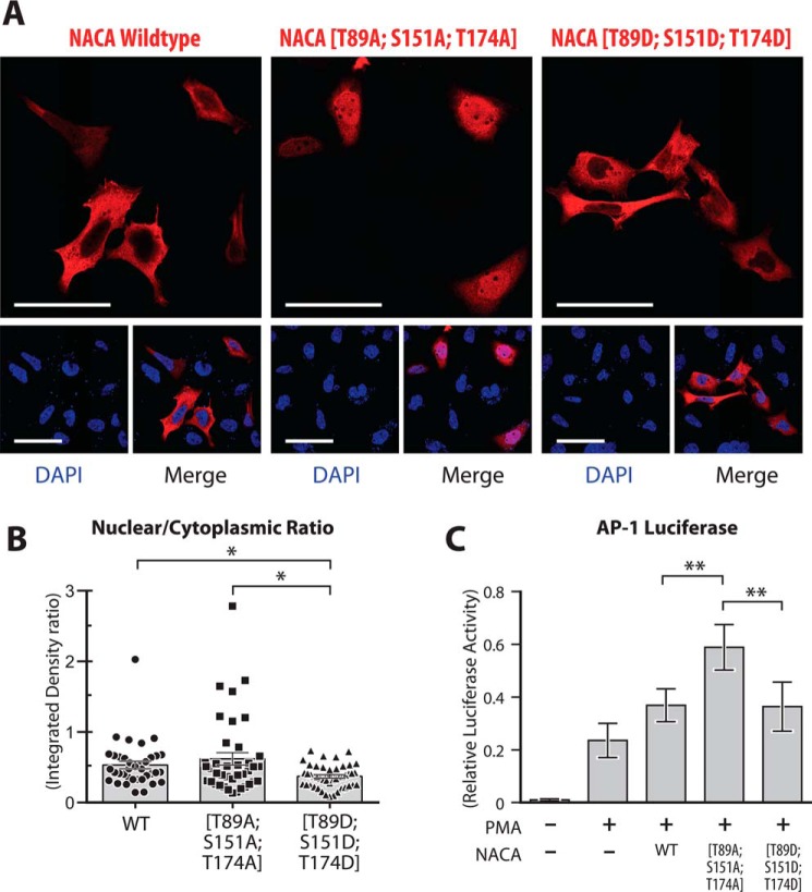
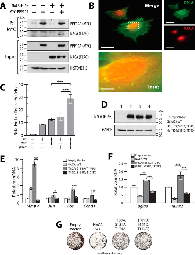
The transcriptional cofactor nascent polypeptide-associated complex and co-regulator α (NACA) regulates osteoblast maturation and activity. NACA functions, at least in part, by binding to Jun proto-oncogene, AP-1 transcription factor subunit (cJUN) and potentiating the transactivation of AP-1 targets such as osteocalcin (Bglap) and matrix metallopeptidase 9 (Mmp9). NACA activity is modulated by phosphorylation carried out by several kinases, but a phosphatase regulating NACA's activity remains to be identified. Here, we used affinity purification with MS in HEK293T cells to isolate NACA complexes and identified protein phosphatase 1 catalytic subunit α (PP1A) as a NACA-associated Ser/Thr phosphatase. NACA interacted with multiple components of the PP1A holoenzyme complex: the PPP1CA catalytic subunit and the regulatory subunits PPP1R9B, PPP1R12A and PPP1R18. MS analysis revealed that NACA co-expression with PPP1CA causes dephosphorylation of NACA at Thr-89, Ser-151, and Thr-174. NACA Ser/Thr-to-alanine variants displayed increased nuclear localization, and NACA dephosphorylation was associated with specific recruitment of novel NACA interactants, such as basic transcription factor 3 (BTF3) and its homolog BTF3L4. NACA and PP1A cooperatively potentiated cJUN transcriptional activity of the AP-1-responsive MMP9-luciferase reporter, which was abolished when Thr-89, Ser-151, or Thr-174 were substituted with phosphomimetic aspartate residues. We confirmed the NACA-PP1A interaction in MC3T3-E1 osteoblastic cells and observed that NACA phosphorylation status at PP1A-sensitive sites is important for the regulation of AP-1 pathway genes and for osteogenic differentiation and matrix mineralization. These results suggest that PP1A dephosphorylates NACA at specific residues, impacting cJUN transcriptional activity and osteoblast differentiation and function.

Keywords: AP1 transcription factor (AP-1); JUN, transcription; PP1A; Protein phosphatase 1 catalytic subunit alpha (PP1A); c-Jun transcription factor; cell differentiation; cell signaling; nascent polypeptide-associated complex and co-regulator alpha (NACA); osteoblast; phosphatase; αNAC.


Conflict of interest statement

The authors declare that they have no conflicts of interest with the contents of this article

Figures

Figure 1.
Figure 1.
PP1A associates with NACA. A, schematic overview of the proteomics approach used to identify NACA-interacting proteins. B, silver-stained SDS-PAGE gel showing expression and purification of NACA protein. C, mass spectrometry (MS/MS) analysis identified numerous NACA-associated proteins, including several components of the PP1A holoenzyme complex. D, validation of interaction of NACA-FLAG with endogenous PP1A complex proteins. HEK293T cells expressing NACA-FLAG or FLAG empty vector were lysed and immunoprecipitated (IP) with anti-FLAG antibody beads and blotted with anti-FLAG antibody or the indicated PP1A-complex antibodies. E, GST-pulldown analysis showing direct interaction between NACA and PPP1CA as well as PPP1R18. GST-NACA fusion protein or GST alone expressed in bacteria (left panel) was incubated with in vitro translated PPP1R9B, PPP1R12A, PPP1R18, and PPP1CA, and then subjected to SDS-PAGE and Western blot analysis (right panel).
Figure 2.
Figure 2.
PP1A dephosphorylates NACA in vitro and in vivo. A, recombinant NACA (arrow) was treated with the indicated recombinant enzymes for 90 min at 30 °C and then analyzed by Western blotting (WB). B, HEK293T cells were transfected with plasmid vectors expressing NACA or the PP1A catalytic unit (PPP1CA). Forty-eight h after transfection, cell lysates were separated by SDS-PAGE and analyzed by Western blotting. In vitro dephosphorylation of NACA (arrow) with recombinant phosphatase enzymes (A) or in vivo co-expression of NACA (B) with PP1A resulted in a faster migrating hypo-phosphorylated form of NACA (arrowhead).
Figure 3.
Figure 3.
PP1A mediates dephosphorylation of NACA at specific residues. A, NACA was immunopurified from HEK293T cells expressing NACA with or without the PP1A catalytic unit (PPP1CA). Posttranslational modifications were identified by LC-tandem MS. B, heat maps indicating the differential abundance of phosphorylation sites detected on NACA in the presence or absence of PPP1CA. Black indicates zero spectral counts and increasing quantities of spectral counts are represented in yellow. C, a representative mass spectra of a NACA phosphopeptide modified at serine 151.
Figure 4.
Figure 4.
NACA dephosphorylation leads to recruitment of novel interactants. A, Venn diagram showing overlap and comparison between the sets of NACA-associated proteins identified by MS. Network analysis and visualization of the identified NACA interactants based on curated protein– interaction databases (GeNets). Interactants unique to PP1A overexpression are delineated within the red rectangle. Closely connected protein clusters are color coded (green, intermediate filaments; purple, cajal bodies; orange, cytoskeleton; blue, mRNA processing; red, transcriptional regulation; interactants that are not part of defined clusters are shown in gray). B, table of the PP1A-dependent NACA interactome. NACA-associated proteins identified by MS in the presence or absence of PPP1CA. C, validation of NACA interaction with BTF3 in a PP1A-dependent manner. HEK293T cells expressing NACA-FLAG and/or MYC-PPP1CA were lysed and immunoprecipitated (IP) with anti-FLAG antibody beads and probed for endogenous BTF3. Overexpression of MYC-PPP1CA induces interaction of NACA and BTF3.
Figure 5.
Figure 5.
PPP1CA and BTF3 functionally cooperate with NACA to regulate cJUN transcription. A, transcriptional activity of a cJUN-responsive MMP9-promoter luciferase (MMP9-Luc) reporter construct in the presence of the indicated expression vectors. B, luciferase activity of a cJUN-responsive construct containing six tandem repeats of the AP-1 binding element (AP1-Luc) in cells transfected with the indicated expression vectors. C, effect of NACA serine/threonine-to-aspartate mutations on cJUN activity on the MMP9-Luc reporter. HEK293T cells were transiently transfected with reporter vectors together with cJUN, NACA, BTF3 and/or PPP1CA expression vectors alone or in combination and relative luciferase activity measured 24 h after transfection. Data are presented as mean ± S.D.; n ≥ 3. **, p < 0.01; ***, p < 0.001; ANOVA with post hoc test. NS, non significant.
Figure 6.
Figure 6.
Regulation of NACA localization by PP1A-sensitive NACA residues. A, HEK293T cells were transiently transfected with WT NACA or NACA mutant constructs and immunostained for NACA with anti-FLAG antibody. B, quantification of NACA distribution in cells treated as in (A) by fluorescence intensity analysis with ImageJ software. C, effect of NACA serine/threonine-to-aspartate or -alanine mutations on AP-1 activity using the AP1-Luc reporter. HEK293T cells were transiently transfected with reporter vectors together with WT NACA or the indicated mutants. Relative luciferase activity was measured 16 h after treatment with 200 nm PMA. Data are presented as mean ± S.D.; n ≥ 3. *, p < 0.05; **, p < 0.01; ANOVA with post hoc test. Magnification bars, 50 μm.
Figure 7.
Figure 7.
NACA and PPP1CA regulate cJUN activity in osteoblast cells. A, interaction of NACA with PPP1CA. MC3T3-E1 cells expressing NACA-FLAG and/or MYC-PPP1CA were lysed and immunoprecipitated (IP) with anti-MYC antibody beads and blotted with the indicated antibodies. B, localization of NACA and PPP1CA in MC3T3-E1 cells was examined by immunofluorescence using antibodies against endogenous NACA (red) and endogenous PPP1CA (green). Areas of colocalization (yellow) were detected in the merged image. C, transcriptional activity of a cJUN-responsive luciferase reporter construct (AP1-Luc) in the presence of the indicated expression vectors in MC3T3-E1 cells. D, immunoblots of whole-cell lysates from MC3T3-E1 cells stably expressing WT NACA or NACA serine/threonine-to-aspartate or -alanine mutations. E, RT-qPCR analysis of AP-1 target gene expression in MC3T3-E1 cells stably expressing NACA or the indicated mutants. F, RT-qPCR analysis of osteoblast differentiation markers after 12 days of differentiation. G, MC3T3-E1 cells differentiated as in (F) were stained for mineral by von Kossa staining. Data are presented as mean ± S.D.; n ≥ 3. *, p < 0.05; ***, p < 0.001; ANOVA with post hoc test. Magnification bars, 50 μm except inset, 9 μm.

References

    1. Chinenov Y., and Kerppola T. K. (2001) Close encounters of many kinds: Fos-Jun interactions that mediate transcription regulatory specificity. Oncogene 20, 2438–2452 10.1038/sj.onc.1204385 - DOI - PubMed
    1. Karin M., Liu Z.-G., and Zandi E. (1997) AP-1 function and regulation. Curr. Opin. Cell Biol. 9, 240–246 10.1016/S0955-0674(97)80068-3 - DOI - PubMed
    1. Shaulian E., and Karin M. (2002) AP-1 as a regulator of cell life and death. Nat. Cell Biol. 4, E131–E136 10.1038/ncb0502-e131 - DOI - PubMed
    1. Wagner E. F., and Eferl R. (2005) Fos/AP-1 proteins in bone and the immune system. Immunol. Rev. 208, 126–140 10.1111/j.0105-2896.2005.00332.x - DOI - PubMed
    1. Bohmann D., Bos T. J., Admon A., Nishimura T., Vogt P. K., and Tjian R. (1987) Human proto-oncogene c-jun encodes a DNA binding protein with structural and functional properties of transcription factor AP-1. Science 238, 1386–1392 10.1126/science.2825349 - DOI - PubMed

Publication types

MeSH terms