. 2019 Apr 4;10(1):1529.
doi: 10.1038/s41467-019-09562-7.

Lower synaptic density is associated with depression severity and network alterations

Affiliations

Lower synaptic density is associated with depression severity and network alterations

Sophie E Holmes et al. Nat Commun. .

Abstract

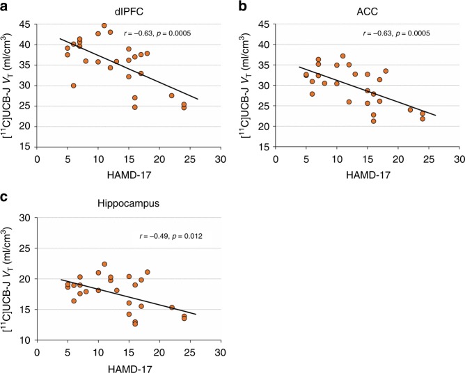
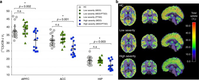
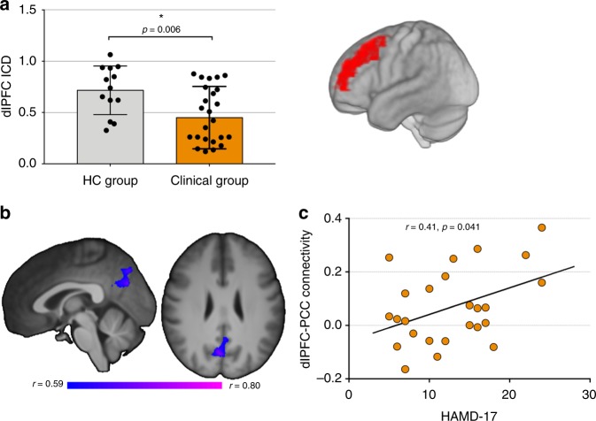
Synaptic loss and deficits in functional connectivity are hypothesized to contribute to symptoms associated with major depressive disorder (MDD) and post-traumatic stress disorder (PTSD). The synaptic vesicle glycoprotein 2A (SV2A) can be used to index the number of nerve terminals, an indirect estimate of synaptic density. Here, we used positron emission tomography (PET) with the SV2A radioligand [11C]UCB-J to examine synaptic density in n = 26 unmedicated individuals with MDD, PTSD, or comorbid MDD/PTSD. The severity of depressive symptoms was inversely correlated with SV2A density, and individuals with high levels of depression showing lower SV2A density compared to healthy controls (n = 21). SV2A density was also associated with aberrant network function, as measured by magnetic resonance imaging (MRI) functional connectivity. This is the first in vivo evidence linking lower synaptic density to network alterations and symptoms of depression. Our findings provide further incentive to evaluate interventions that restore synaptic connections to treat depression.


Conflict of interest statement

Dr. Krystal acknowledges the following relevant financial interests. He is a co-sponsor of a patent for the intranasal administration of ketamine for the treatment of depression that was licensed by Janssen Pharmaceuticals, the maker of s-ketamine. He has a patent related to the use of riluzole to treat anxiety disorders that was licensed by Biohaven Medical Sciences. He has stock or stock options in Biohaven Medical Sciences, ARett Pharmaceuticals, Blackthorn Therapeutics, and Luc Therapeutics. He consults broadly to the pharmaceutical industry, but his annual income over the past year did not exceed $5,000 for any organization. He receives over $5,000 in income from the Society of Biological Psychiatry for editing the journal Biological Psychiatry. He has fiduciary responsibility for the International College of Neuropsychopharmacology as president of this organization. The remaining authors declare no competing interests.

Figures

Fig. 1
Fig. 1
Correlations between SV2A density and severity of depressive symptoms in the full clinical sample Correlations between [11C]UCB-J VT and HAMD-17 scores in dlPFC (a), ACC (b) and hippocampus (c) across all clinical subjects (n = 26). Correlations were computed using Pearson’s r. dlPFC: dorsolateral prefrontal cortex, ACC: anterior cingulate cortex
Fig. 2
Fig. 2
Lower SV2A density in individuals with high severity depressive symptoms vs. HC subjects. a [11C]UCB-J VT in the dlPFC, ACC, and hippocampus across groups. The low severity group (in green; n = 14) consisted of participants with HAMD-17 scores <14 and the high severity group (in blue; n = 12) consisted of participants with HAMD-17 scores ≥14. Clinical subgroups are represented by different symbols; the low severity group consisted of MDD (n = 5), PTSD (n = 5) and MDD/PTSD (n = 4) subjects; the high severity group consisted of MDD (n = 6) and MDD/PTSD (n = 6) subjects. Group differences were assessed using MANOVA. Error bars represent standard deviation. b Representative axial, coronal, and sagittal parametric images of [11C]UCB-J PET (VT) scans registered to MR images in MNI space from a HC participant (top), low severity subject (middle) and high severity subject (bottom). Color bar represents VT. dlPFC: dorsolateral prefrontal cortex: dlPFC, ACC:anterior cingulate cortex
Fig. 3
Fig. 3
Functional connectivity and combined connectivity/PET results. a Significantly lower dlPFC ICD connectivity in clinical (n = 26) vs. HC group (n = 13). Error bars represent standard deviation. dlPFC region displayed on right, subsequently used as seed. b Negative correlation between dlPFC-PCC connectivity and dlPFC SV2A density in the clinical group. Significant voxels represent those voxels whose connectivity with the dlPFC negatively correlates with [11C]UCB-J VT in the dlPFC. c Positive correlation between dlPFC-PCC connectivity and severity of depressive symptoms. Correlations were computed using Pearson’s r. dlPFC: dorsolateral prefrontal cortex, PCC: posterior cingulate cortex, ICD: intrinsic connectivity distribution
Fig. 4
Fig. 4
Stronger ACC-hippocampus correlation in SV2A density in high severity vs. low severity clinical groups. Differences in correlations between ROIs were assessed using Fisher’s r to z transformation. ACC: anterior cingulate cortex, HIP: hippocampus
Fig. 5
Fig. 5
Relationship between SV2A density and functional connectivity, and cognitive function. a Lower performance on verbal memory task (International Shopping List-Delayed Recall) in clinical (n = 26) vs HC (n = 15) group. Error bars represent standard deviation. b Significant positive correlation between [11C]UCB-J VT in verbal memory across clinical subjects. c Trend level correlation between dlPFC SV2A density and verbal memory. d Lower performance on working memory task (one-back) in high severity clinical subjects (n = 12) vs. HC (n = 15). e Positive correlation between dlPFC ICD connectivity and working memory in high severity clinical subjects. f Negative correlation between dlPFC-PCC connectivity and working memory in high severity clinical subjects, suggesting that the greater the connectivity between core nodes of two typically anticorrelated networks (default mode and central executive networks), the greater the impairment in working memory. Correlations were assessed using Pearson’s r. dlPFC: dorsolateral prefrontal cortex, PCC: posterior cingulate cortex, ICD: intrinsic connectivity distribution

References

    1. Duman RS, Aghajanian GK, Sanacora G, Krystal JH. Synaptic plasticity and depression: new insights from stress and rapid-acting antidepressants. Nat. Med. 2016;22:238. doi: 10.1038/nm.4050. - DOI - PMC - PubMed
    1. Kessler RC, et al. Lifetime prevalence and age-of-onset distributions of DSM-IV disorders in the National Comorbidity Survey Replication. Arch. Gen. Psychiatry. 2005;62:593–602. doi: 10.1001/archpsyc.62.6.593. - DOI - PubMed
    1. Goodkind M, et al. Identification of a common neurobiological substrate for mental illness. JAMA Psychiatry. 2015;72:305–315. doi: 10.1001/jamapsychiatry.2014.2206. - DOI - PMC - PubMed
    1. Kang HJ, et al. Decreased expression of synapse-related genes and loss of synapses in major depressive disorder. Nat. Med. 2012;18:1413. doi: 10.1038/nm.2886. - DOI - PMC - PubMed
    1. Feyissa AM, Chandran A, Stockmeier CA, Karolewicz B. Reduced levels of NR2A and NR2B subunits of NMDA receptor and PSD-95 in the prefrontal cortex in major depression. Progress. Neuro-Psychopharmacol. Biol. Psychiatry. 2009;33:70–75. doi: 10.1016/j.pnpbp.2008.10.005. - DOI - PMC - PubMed

Publication types