Review
. 2019 Jul;20(7):389-405.
doi: 10.1038/s41580-019-0116-4.

Establishing and regulating the composition of cilia for signal transduction

Affiliations
Review

Establishing and regulating the composition of cilia for signal transduction

Maxence V Nachury et al. Nat Rev Mol Cell Biol. 2019 Jul.

Abstract

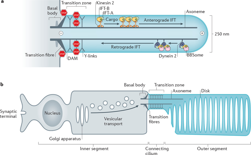
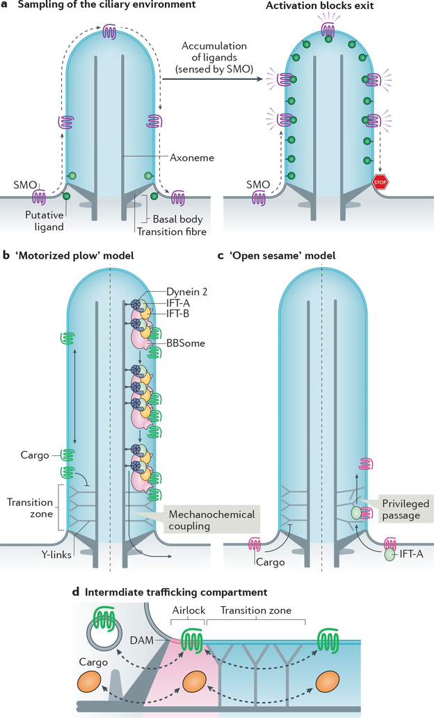
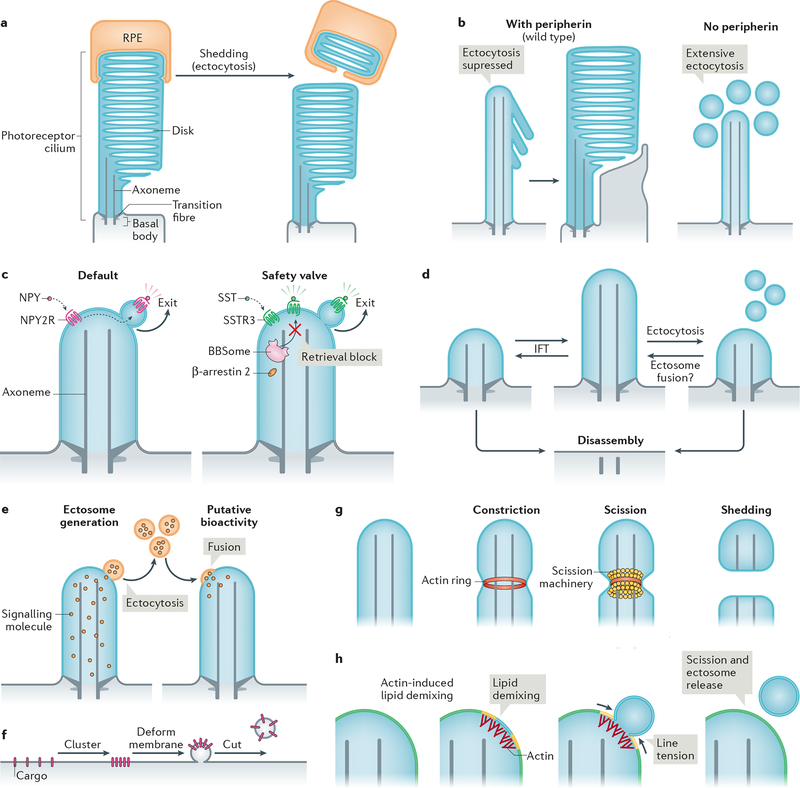
The primary cilium is a hair-like surface-exposed organelle of the eukaryotic cell that decodes a variety of signals - such as odorants, light and Hedgehog morphogens - by altering the local concentrations and activities of signalling proteins. Signalling within the cilium is conveyed through a diverse array of second messengers, including conventional signalling molecules (such as cAMP) and some unusual intermediates (such as sterols). Diffusion barriers at the ciliary base establish the unique composition of this signalling compartment, and cilia adapt their proteome to signalling demands through regulated protein trafficking. Much progress has been made on the molecular understanding of regulated ciliary trafficking, which encompasses not only exchanges between the cilium and the rest of the cell but also the shedding of signalling factors into extracellular vesicles.


Conflict of interest statement

Competing interests

The authors declare no competing interests.

Figures

Figure 1|
Figure 1|. Organization of primary cilia and photoreceptors.
a| Diagram of a primary cilium. The basal body consists of 9 triplets of microtubules and is connected to the plasma membrane by the transition fibres. The axoneme is formed by 9 doublets of microtubules (only 2 are shown). At the base of the axoneme is the transition zone, which is connected to the ciliary membrane by Y-links. Microtubule motors and three intraflagellar transport (IFT) complexes (Box 3) assemble into IFT trains that move ciliary cargoes move along the axoneme.. Kinesin-II mediates anterograde IFT and cytoplasmic dynein 2 retrograde IFT. The DAM (distal appendage matrix) –positioned between the transiton fibres together with the transition zone separate the cilium from the rest of the cell by blocking (STOP signs) lateral diffusion of membrane proteins and diffusion of soluble proteins. Plus (+) and minus (–) mark the orientation of the microtubules in the axoneme. b| Diagram of a photoreceptor neuron. The outer segment (equivalent of the cilium shaft) comprises hundreds of membrane disks that each concentrates about 1 million molecules of rhodopsin. All proteins required for phototransduction are produced in the inner segment, transported through the transition zone (termed ‘connecting cilium’), and are injected into the outer segment.
Figure 2|
Figure 2|. Transport processes that shape the ciliary environment.
a| Local supply of ciliary molecules. Left: ciliary calcium channels allow local influx of Ca2+ into cilia, which then passively leaks into the cytoplasm (dashed arrows). Middle: adenylyl cyclases generate cAMP in cilia, which then passively leaks into the cytoplasm (dashed arrows). Right: phosphatidylinositol (PtdIns) 5-phosphatase INPP5E in the ciliary membrane converts PtdIns(4,5)P2 into PtdIns(4)P. Dashed arrows indicate lipid exchange between cilia and plasma membrane (assuming that there is no diffusion barrier for lipids). b| A diffusion-to-capture mechanism can enrich certain ciliary proteins. One example is microtubule plus-end binding protein EB1. c| Some membrane proteins, such as Chlamydomonas reinhardtii adhesion receptor SAG1 can enter cilia and accumulate at the ciliary base in the absence of intraflagellar transport (IFT) proteins. How this occurs or whether there are additional factors required is unknown (question mark). Similarly, tubulin dimers enter cilia by passive diffusion (dashed arrows) but require consumption of energy (solid arrow) to efficiently reach the growing ends of axonemal microtubules by IFT. d| Import of certain G-protein coupled receptors (GPCRs) by the TULP3–IFT-A complex. IFT-A recognizes ciliary targeting signals within GPCRs, and unloads its cargoes upon hydrolysis of PtdIns(4,5)P2 to PtdIns(4)P in the cilium. This ‘injection’ into the cilium may not require mechanochemical coupling of kinesin-II. e| Import of prenylated ciliary proteins is facilitated by specific chaperones that shield the hydrophobic prenyl groups. Exemplified here is INPP5E, whose farnesyl group (zigzag) is chaperoned by PDE6δ. Upon entry of the INPP5E–PDE6δ complex into cilia, the binding of ARL3–GTP to PDE6δ triggers the release of INPP5E, which becomes membrane-associated. GTP loading on ARL3 is promoted by ciliary protein ARL13B due to its guanine nucleotide exchange factor (GEF) activity, which ensures high levels of ARL3–GTP in cilia. RP2 at the transition zone functions as a GTPase-activating protein (GAP) for ARL3, ensuring that ARL3 is GDP-bound after exiting cilia. This creates a gradient of ARL3–GTP, with ARL3–GTP — and consequently INPP5E release — being restricted to the ciliary shaft. f| Hierarchy of ciliary localizations during protein targeting to cilia. Shown are factors required to allow for an enrichment of IFT-A cargos in cilia. ARL13B in cilia ensures GTP loading of ARL3, which is required for INPP5E localization to cilia. INPP5E hydrolyzes PtdIns(4,5)P2 to PtdIns(4)P in cilia, which allows cargo release from TULP3–IFT-A. How ARL13B is enriched in cilia is currently unknown. g| Passive influx of molecules can be overcome by active removal, exemplified by the BBSome-dependent retrieval of phospholipase D (PLD). h| Activated GPCRs are recognized by the β-arrestin 2, followed by capture by nascent BBSome-containing IFT trains at the ciliary tip, processive retrograde IFT and ultimately transition zone crossing. This may be followed by either GPCR endocytosis or lateral diffusion into the plasma membrane, both outcomes leading to receptor retrieval from the cilium into the cell.
Figure 3|
Figure 3|. Models of transition zone crossing and of the intermediate compartment.
a| Constitutive passage through the cilium enables Smoothened (SMO) to continuously sample the environment of the cilium. Elevation of inner leaflet cholesterol (see Box 2) or other second messengers leads to the activation of SMO and blockage of exit to result in ciliary accumulation of activated SMO and activation of downstream signalling. b|c| Membrane proteins cannot cross the transition zone unassisted. According to the ‘motorized plow’ model (b), dynein 2 utilizes the energy of ATP to generate microtubule-based motion and drags retrograde intraflagellar transport (IFT) trains and their attached cargoes through the transition zone (right). According to the ‘open sesame’ model (c), cargo binding to import factors (exemplified by IFT-A) allows privileged passage of the transition zone in a motor-independent manner (right). In this model, the transport factors locally remodel the transition zone to allow passage. Note that the models may apply to either trafficking directions. d| Increasing evidence points towards the existence of an intermediate trafficking compartment at the ciliary base that functions as an ‘airlock’ proximal to the transition zone and may thus represent a checkpoint for ciliary import and export. The proximal boundary of the airlock is likely to correspond to the distal appendage matrix (DAM) located at the membrane between transition fibres.
Figure 4|
Figure 4|. Modalities and mechanisms of ciliary ectocytosis.
a| The tips of photoreceptor outer segments are shed daily to remove aged rhodopsin molecules. Shed material is phagocytosed by retinal pigmented epithelial (RPE) cells (and recycled back to photoreceptors; not shown) (left). Ectocytosis also negatively regulates outer segment morphogenesis: the presence of disk protein peripherin blocks ectocytosis and enables retention of ciliary components to form disks (right). b| Upon activation, some G protein-coupled receptors (GPCRs) are removed from cilia by ectocytosis. For example, neuropeptide Y receptor type 2 (NPY2R) uses ectocytosis as a primary mechanism for cilia exit (left). Activated somatostatin receptor 3 (SSTR3) becomes ectocytosed when its BBSome-based retrieval (see Fig. 3b) is blocked (right). NPY, neuropeptide Y, SST, somatostatin. c| Intraflagellar transport (IFT) elongates or shrinks the cilium by exchanging material with the rest of the cell. Ectocytosis may serve as an alternative mechanism of ciliary length homeostasis: shedding of fragments of cilia from the tip by ectocytosis reduces ciliary length, whereas the hypothetical fusion of extracellular vesicles with the cilium may increase cilia length. d| Cilia package signalling molecules into ectosomes. These may be received by other cells to confer putative bioactivity. As exemplified in Chlamydomonas reinhardtii, recipient cells may receive extracellular vesicles via their cilia; incorporation via the plasma membrane is also conceivable. e| Basic steps of extracellular vesicle formation: clustering of cargoes is followed by membrane deformation and finally, a forming vesicle is released from the donor membrane by scission. f| Actin may serve to constrict the diameter of the cilium by forming a contractile ring prior to scission driven by an additional scission machinery, such as ESCRT-III-based complexes. g| Model of how actin may function in ectosome budding and scission. For simplicity, the ciliary membrane is depicted as consisting of two lipid species (yellow and blue, which blend to green). Actin polymerization in cilia causes local lipid demixing. Line tension at the interface between lipid phases ensues, which is resolved by budding and scission of the de-mixed lipids, releasing a vesicle.

References

    1. Reiter JF & Leroux MR Genes and molecular pathways underpinning ciliopathies. Nat. Rev. Mol. Cell Biol 18, 533–547 (2017). - PMC - PubMed
    1. de Robertis E Electron microscope observations on the submicroscopic organization of the retinal rods. The Journal of biophysical and biochemical cytology 2, 319–330 (1956). - PMC - PubMed
    1. Bloom G Studies on the olfactory epithelium of the frog and the toad with the aid of light and electron microscopy. Zeitschrift für Zellforschung und Mikroskopische Anatomie 41, 89–100 (1954). - PubMed
    1. Huangfu D et al. Hedgehog signalling in the mouse requires intraflagellar transport proteins. Nature 426, 83–7 (2003). - PubMed
    1. Gerdes JM et al. Disruption of the basal body compromises proteasomal function and perturbs intracellular Wnt response. Nature Genetics 39, 1350–1360 (2007). - PubMed

Publication types