. 2019 Mar 15:13:213.
doi: 10.3389/fnins.2019.00213. eCollection 2019.

Brain Microglial Activation in Chronic Pain-Associated Affective Disorder

Affiliations

Brain Microglial Activation in Chronic Pain-Associated Affective Disorder

Ellane Eda Barcelon et al. Front Neurosci. .

Abstract

A growing body of evidence from both clinical and animal studies indicates that chronic neuropathic pain is associated with comorbid affective disorders. Spinal cord microglial activation is involved in nerve injury-induced pain hypersensitivity characterizing neuropathic pain. However, there is a lack of thorough assessments of microglial activation in the brain after nerve injury. In the present study, we characterized microglial activation in brain sub-regions of CX3CR1GFP/+ mice after chronic constriction injury (CCI) of the sciatic nerve, including observations at delayed time points when affective brain dysfunctions such as depressive-like behaviors typically develop. Mice manifested chronic mechanical hypersensitivity immediately after CCI and developed depressive-like behaviors 8 weeks post-injury. Concurrently, significant increases of soma size and microglial cell number were observed in the medial prefrontal cortex (mPFC), hippocampus, and amygdala 8 weeks post-injury. Transcripts of CD11b, and TNF-α, genes associated with microglial activation or depressive-like behaviors, are correspondingly upregulated in these brain areas. Our results demonstrate that microglia are activated in specific brain sub-regions after CCI at delayed time points and imply that brain microglial activation plays a role in chronic pain-associated affective disorders.

Keywords: TNF-α; brain microglia; chronic pain; depression; microglial activation.


Figures

FIGURE 1
FIGURE 1
CCI-induced mice developed depressive-like behaviors. (A) Experimental timeline denoting weeks of behavior assessment. Bottom: CCI showing ligation of the sciatic nerve. (B) Mechanical allodynia tests were used to determine paw withdrawal thresholds before surgery (BL: baseline) and significant decreases of the paw withdrawal threshold 1, 4, and 8 weeks post-injury in CCI mice (n = 5, mean ± SEM, ∗∗∗p < 0.001, ∗∗p < 0.01). (C) TST and (D) FST showed significant increases of immobility time in the CCI group compared to the sham group (n = 5, mean ± SEM, p < 0.05) indicating that depressive-like behaviors were induced 8 weeks post-injury. (E) Locomotor activity assessed in terms of total distance traveled in an open field test (OFT), showed no significant difference between groups (n = 5).
FIGURE 2
FIGURE 2
CCI-induced mice showed altered microglial morphology in the mPFC and amygdala at delayed time points. Representative images of (A) mPFC and a high magnification image at 8 weeks (inset) are shown. (B) Quantification of microglial cell number per square millimeter at 1, 4, and 8 weeks (n = 5, 3–4 tissue sections per animal, mean ± SEM, p < 0.05, ∗∗p < 0.01) along with microglial soma area 8 weeks post-injury (n = 5, 3–4 tissue samples per section, 40 microglial cells, mean ± SEM, ∗∗p < 0.01) showing significant increases at 8 weeks in the CCI group compared to the sham group. Similarly, (C) representative images of the amygdala at 1, 4, and 8 weeks after injury, and a higher magnification image taken at 8 weeks (inset) are shown along with (D) microglial cell number and soma area quantification at 1, 4, and 8 weeks post-injury. Scale bar: 100 μm. Inset scale bar: 100 μm.
FIGURE 3
FIGURE 3
CCI-induced mice showed altered microglial morphology in hippocampal layers at delayed time points. (A) Representative images of the hippocampus and images taken at 1, 4, and 8 weeks post-injury under high magnification (inset) are shown. (B) Microglial cell number in the hippocampus shows significant increases at 4 and 8 weeks (n = 5, 3–4 tissue sections per animal, mean ± SEM, p < 0.05, ∗∗p < 0.01) along with significant increases of the microglia soma area (n = 5, 3–4 tissue samples per section, 40 microglial cells, mean ± SEM, ∗∗p < 0.01). (C) Hippocampal layers, namely the stratum oriens (SO), stratum lacunosum moleculare (SLM), molecular layer (ML), and hilus (H) were demonstrated. (D) Hippocampal layer-specific microglial cell numbers were assessed at 1, 4, and 8 weeks post-injury and demonstrated significant differences in the CCI group compared to the sham group (n = 5, 3–4 tissue sections per animal, mean ± SEM, p < 0.05, ∗∗p < 0.01, ∗∗∗p < 0.001). Scale bar: 100 μm. Inset scale bar: 100 μm.
FIGURE 4
FIGURE 4
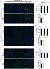
CCI did not show obvious changes of microglial morphology in the nucleus accumbens, periaqueductal gray, or habenula at any of the time points investigated. Representative images of the (A) nucleus accumbens, (C) periaqueductal gray, (E) habenula, and a high magnification image taken at 8 weeks post-injury (inset) are shown, along with (B, D, F) the areas of quantification of microglial cell number (n = 5, 3–4 tissue sections per animal, mean ± SEM) at 1, 4, and 8 weeks and soma area (n = 5, 3–4 tissue samples per section, 40 microglial cells, mean ± SEM) at 8 weeks post-injury showing no significant differences between the CCI group and sham group. Scale bar: 100 μm. Inset scale bar: 100 μm.
FIGURE 5
FIGURE 5
CCI-induced neuropathic pain induced changes in gene expression related to microglial activation and depression in the mPFC, amygdala (Amy), and hippocampus (Hipp) at delayed time points (8 weeks) post-injury. qRT-PCR showed significantly increased expression of (A) CD11b, (B) TMEM119, (C) P2RX7 (D) P2RY12, and (E) TNF-α in brain areas observed post-injury (n = 3∼6, mean ± SEM, p < 0.05, ∗∗p < 0.01). However, qRT-PCR showed no significant differences in (F) iNOS and (G) IL-6 expression levels between CCI and sham groups post-injury (n = 3∼6).

References

    1. Beggs S., Salter M. W. (2010). Microglia–neuronal signalling in neuropathic pain hypersensitivity 2.0. Curr. Opin. Neurobiol. 20 474–480. 10.1016/j.conb.2010.08.005 - DOI - PMC - PubMed
    1. Beggs S., Trang T., Salter M. W. (2012). P2x4r+ microglia drive neuropathic pain. Nat. Neurosci. 15 1068–1073. 10.1038/nn.3155 - DOI - PMC - PubMed
    1. Bennett G. J., Xie Y. K. (1988). A peripheral mononeuropathy in rat that produces disorders of pain sensation like those seen in man. Pain 33 87–107. 10.1016/0304-3959(88)90209-6 - DOI - PubMed
    1. Bennett M. L., Bennett F. C., Liddelow S. A., Ajami B., Zamanian J. L., Fernhoff N. B., et al. (2016). New tools for studying microglia in the mouse and human CNS. Proc. Natl. Acad. Sci. U.S.A. 113 E1738–E1746. 10.1073/pnas.1525528113 - DOI - PMC - PubMed
    1. Blank T., Prinz M. (2013). Microglia as modulators of cognition and neuropsychiatric disorders. Glia 61 62–70. 10.1002/glia.22372 - DOI - PubMed