. 2019 Mar 20:9:171.
doi: 10.3389/fonc.2019.00171. eCollection 2019.

Lysosomal Pathways and Autophagy Distinctively Control Endothelial Cell Behavior to Affect Tumor Vasculature

Affiliations

Lysosomal Pathways and Autophagy Distinctively Control Endothelial Cell Behavior to Affect Tumor Vasculature

Marco B Schaaf et al. Front Oncol. .

Abstract

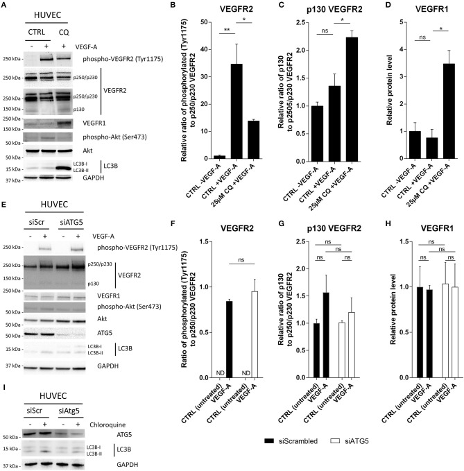
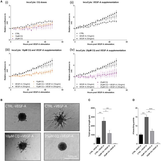
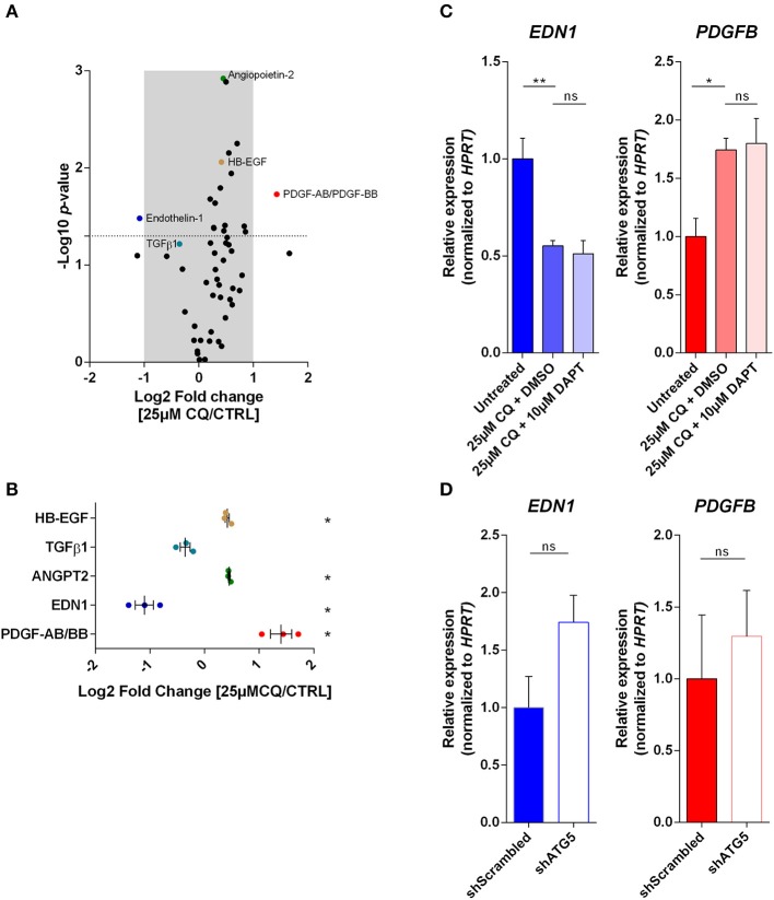
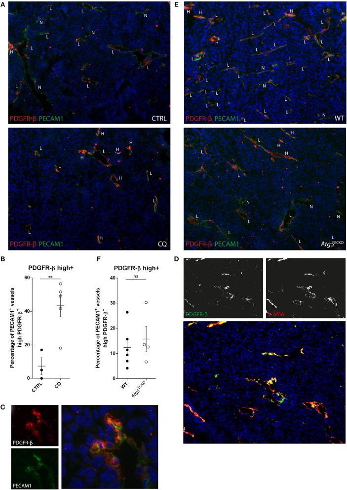
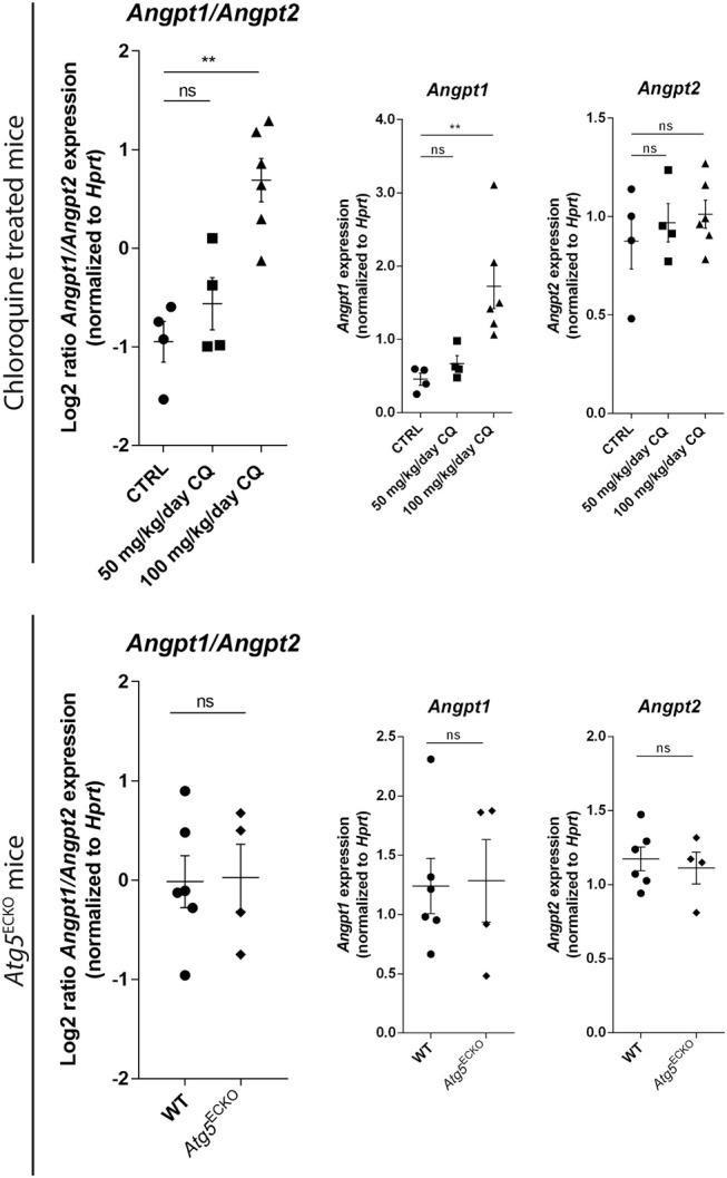
Cancer cell-stromal cell crosstalk is orchestrated by a plethora of ligand-receptor interactions generating a tumor microenvironment (TME) which favors tumor growth. The high pro-angiogenic nature of the TME perpetuates the chaotic network of structurally immature, low pericyte-covered vessels characteristic of the tumor vasculature. We previously demonstrated that chloroquine (CQ) -a lysosomotropic agent used as first-generation autophagy blocker in clinical trials- induced tumor vessel normalization and reduced tumor hypoxia. CQ improved both vessel structure and maturation, whereas the conditional knockout of the crucial autophagy gene Atg5 in endothelial cells (ECs) did not, thus highlighting a potential differential role for EC-associated autophagy and the lysosomes in pathological tumor angiogenesis. However, how CQ or ATG5-deficiency in ECs affect angiogenic signals regulating EC-pericyte interface and therefore vessel maturation, remains unknown. Here, we show that in ECs CQ constrained VEGF-A-mediated VEGF receptor (VEGFR)2 phosphorylation, a driver of angiogenic signaling. In the presence of CQ we observed increased expression of the decoy receptor VEGFR1 and of a lower molecular weight form of VEGFR2, suggesting receptor cleavage. Consequently, VEGF-A-driven EC spheroid sprouting was reduced by CQ treatment. Furthermore, CQ significantly affected the transcription and secretion of platelet-derived growth factor (PDGF)-AB/BB (upregulated) and Endothelin-1 (EDN1, downregulated), both modulators of perivascular cell (PC) behavior. In contrast, silencing of ATG5 in ECs had no effect on VEGFR2 to VEGFR1 ratio nor on PDGFB and EDN1 expression. Accordingly, mice harboring B16F10 melanoma tumors treated with CQ, displayed both an increased number of αSMA+ PCs covering tumor vessels and co-expressed PDGF receptor-β, enabling PDGF ligand dependent recruitment. Moreover, upon CQ treatment the tumoral expression of angiopoietin-1 (Angpt1), which retains mural cells, and induces vessel stabilization by binding to the EC-localized cognate receptor (TIE2), was increased thus supporting the vessel normalization function of CQ. These features associated with improved tumor vasculature were not phenocopied by the specific deletion of Atg5 in ECs. In conclusion, this study further unravels endothelial cell autonomous and non-autonomous mechanisms by which CQ "normalizes" the intercellular communication in the tumor vasculature independent of autophagy.

Keywords: Angiopoietin1; Autophagy; PDGFR-β; VEGF/VEGFR-axis; intercellular crosstalk; tumor endothelial cells.


Figures

Figure 1
Figure 1
CQ treatment, but not ATG5 deficiency, reduces VEGFR2 phosphorylation after VEGF-A stimulation. HUVECs were either (A–D) pretreated with CQ or (E-I) siRNA against ATG5 (siATG5) and treated with 50 ng/ml VEGF-A where indicated. Cells were treated with non-targeting siRNA (siScrambled; siScr) as control. (A,E,I) Cell lysates were analyzed by immunoblotting. (B,F) Graphs display (relative) band intensity ratio of phosphorylated or (C,G) p130 VEGFR2 to p250/p230 VEGFR2. (D,G) Bar graphs display VEGFR1 intensity normalized to GAPDH. (I) HUVECs were cultured in presence or absence of 25 μM CQ for 2 h. A short-term exposure was opted to minimalize any secondary effects on autophagy flux due to CQ treatment. N.D., not detected. (B–D,F–H), n = 3, mean ± SEM.
Figure 2
Figure 2
CQ reduces proliferation and spheroid sprouting after VEGF-A stimulation. HUVEC proliferation was assessed based on confluency measurements in an IncuCyte imaging system. (A) From start of measurements at t = 0 (the moment of VEGF-A supplementation with indicated doses) images were taken every 2 h up to 48 h. Graphs display confluency results of the indicated conditions (n = 4). At t = 48 h: (ii) p-value < 0.05 for CTRL vs. VEGF-A (50 ng/ml). (iii) p-value < 0.05 for VEGF-A (50 ng/ml) vs. 10 μM CQ + VEGF-A (50 ng/ml). (iv) p-value < 0.01 for VEGF-A (50 ng/ml) vs. 25 μM CQ + VEGF-A (50 ng/ml). (B–D) HUVEC spheroids exposed to 50 ng/ml VEGF-A in presence or absence of 25 μM CQ were analyzed for cumulative sprout length and number of branching points (n = 5–17). (A,C,D), mean ± SEM.
Figure 3
Figure 3
Autophagy and the endo-lysosomal pathway in ECs differentially modulate the secretion and expression of factors involved in EC-PC interaction. (A) Cell culture supernatant of 25 μM CQ-treated and untreated HUVECs was analyzed with a Proteome Profiler Human Angiogenesis Antibody array. Volcano plot indicates the fold change and p-value of the dot densitometry (n = 3). (B) Fold change of selected proteins involved in EC-PC recruitment or differentiation are plotted with each dot representing an individual experiment. (C,D) PDGFB and EDN1 gene expression was analyzed in HUVECs treated with CQ, DAPT or DMSO (vehicle control for DAPT) (n = 3), or, HUVECs expressing non-targeting (shScrambled) or ATG5-targeting (shATG5) shRNA (n = 5). (B–D), mean ± SEM.
Figure 4
Figure 4
CQ treatment increases tumor vessel coverage of PDGFR-β+ cells, but not by endothelial specific Atg5 knockout. (A,C,E) B16F10 melanoma tissue sections were analyzed for PDGFR-β (red) and PECAM1 (green) expression by immunohistochemistry. B16F10 melanomas were harvested from CQ-treated (100 mg/kg/day), Atg5ECKO mice or their respective controls. DAPI was used as counterstaining for nuclei. PECAM1+ vessels were scored negative (N), low (L), or high (H) for PDGFR-β. (B,F) In the graphs, each dot represents the average of at least five images made from a tumor section at 10x magnification. (C) Representative image of a cross-sectioned vessel in a B16F10 melanoma of a CQ (100 mg/kg/day) treated mouse. (D) Representative image of B16F10 melanoma tissue section stained for PDGFR-β (green), αSMA (red), and nuclei (blue). (B,F) mean ± SEM.
Figure 5
Figure 5
Gene expression analyses in tumors validate improved EC-PC interaction. B16F10 melanomas were harvested from CQ-treated [50 or 100 mg/kg/day), Atg5ECKO and their designated control mice (untreated (CTRL) or wild type (WT), respectively]. Lysates were analyzed for the abundance of Angpt1, Angpt2, and Hprt mRNA. Results are plotted in graphs displaying Log2(Angpt1/Angpt2) and abundance of individual gene transcripts. Each dot represents an individual mouse. Mean ± SEM.

References

    1. Welti J, Loges S, Dimmeler S, Carmeliet P. Recent molecular discoveries in angiogenesis and antiangiogenic therapies in cancer. J Clin Invest. (2013) 123:3190–200. 10.1172/JCI70212 - DOI - PMC - PubMed
    1. Carmeliet P, Jain RK. Molecular mechanisms and clinical applications of angiogenesis. Nature. (2011) 473:298–307. 10.1038/nature10144 - DOI - PMC - PubMed
    1. Schaaf MB, Garg AD, Agostinis P. Defining the role of the tumor vasculature in antitumor immunity and immunotherapy. Cell Death Dis. (2018) 9:115. 10.1038/s41419-017-0061-0 - DOI - PMC - PubMed
    1. Blanco R, Gerhardt H. VEGF and Notch in tip and stalk cell selection. Cold Spring Harbor Perspect Med. (2013) 3:a006569. 10.1101/cshperspect.a006569 - DOI - PMC - PubMed
    1. Gerhardt H, Golding M, Fruttiger M, Ruhrberg C, Lundkvist A, Abramsson A, et al. . VEGF guides angiogenic sprouting utilizing endothelial tip cell filopodia. J Cell Biol. (2003) 161:1163–77. 10.1083/jcb.200302047 - DOI - PMC - PubMed