Review
. 2019 Mar 20:7:41.
doi: 10.3389/fcell.2019.00041. eCollection 2019.

Autosomal Dominant Leukodystrophy: A Disease of the Nuclear Lamina

Affiliations
Review

Autosomal Dominant Leukodystrophy: A Disease of the Nuclear Lamina

Quasar S Padiath. Front Cell Dev Biol. .

Abstract

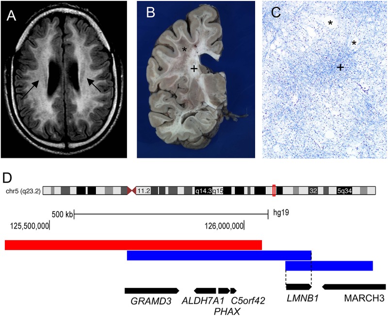
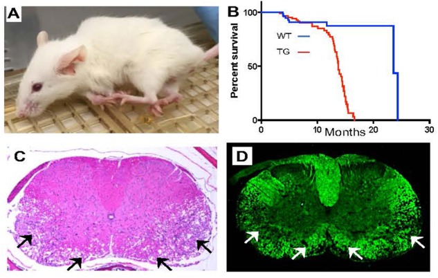
The nuclear lamina is a fibrous meshwork of proteins found adjacent to the inner nuclear membrane that plays a critical role in the maintenance of nuclear architecture. Made up of A and B type lamins, the nuclear lamina has recently been shown to contribute to numerous cellular functions such as chromatin organization, DNA replication, cellular proliferation, senescence, and aging. While at least a dozen disorders are associated with LMNA, the focus of this review is Autosomal Dominant Leukodystrophy (ADLD), the only disease associated with the lamin B1 gene (LMNB1). ADLD is a fatal, adult onset CNS demyelinating disorder that is caused by either genomic duplications involving LMNB1 or deletions upstream of the gene. Both mutation types result in increased LMNB1 gene expression. How the increased levels of this widely expressed nuclear structural component results a phenotype as specific as demyelination is a great mystery. This review summarizes what is currently known about the disease and describes recent work using animal and cell culture models that have provided critical insights into ADLD pathological mechanisms. The delineation of these pathways provides a fascinating glimpse into entirely novel roles for the nuclear lamina and will be critical for the identification of therapies for this fatal disease.

Keywords: chromatin; lamin B1; leukodystrophy; lipid synthesis; myelin; nuclear lamina; nuclear structure.


Figures

FIGURE 1
FIGURE 1
Genomic rearrangements involving the LMNB1 gene cause the demyelinating disorder Autosomal Dominant Leukodystrophy (ADLD). (A) ADLD patient MRI (Fluid-attenuated inversion recovery sequence) reveals white matter hyperintensities indicating myelin pathology (marked by arrows). (B) ADLD patient brain showing patchy areas of myelin loss marked by asterisk (). Plus sign (+) indicates normal myelin. (C) Histopathological analysis of ADLD brain section using Luxol Fast Blue, a myelin stain, exhibits areas of pale staining and vacuolar demyelination, marked by asterisks (). Plus sign (+) indicates normal myelin staining. (D) Genomic region on chromosome 5q23.2 that contains the LMNB1 gene. Blue bars indicate genomic duplications from two individual patients that have centromeric and telomeric junctions closest to the LMNB1 gene that allow the identification of the minimal critical region required for disease causation (dashed line). Red bar indicates the deletion upstream of the LMNB1 gene responsible for a variant ADLD phenotype reported in a single family. Data for (D) is modified from Giorgio et al. (2015). Black bars indicate the location of genes encompassed by the genomic rearrangements and the direction of transcription.
FIGURE 2
FIGURE 2
Mouse models overexpressing LMNB1 recapitulate ADLD phenotypes. (A) Transgenic mice where LMNB1 is targeted to oligodendrocytes show progressive age dependent muscle wasting, kyphosis, limb paralysis and (B) significantly reduced lifespan. Histopathological analysis of spinal cord sections from transgenic mice reveal severe vacuolar degeneration of myelinated regions (arrows). (C) Hematoxylin and Eosin staining. (D) Fluoromyelin (a myelin specific stain) staining [Reproduced with permission from Rolyan et al. (2015)].

References

    1. Alturkustani M., Sharma M., Hammond R., Ang L. C. (2013). Adult-onset leukodystrophy: review of 3 clinicopathologic phenotypes and a proposed classification. J. Neuropathol. Exp. Neurol. 72 1090–1103. 10.1097/NEN.0000000000000008 - DOI - PubMed
    1. Barascu A., Le Chalony C., Pennarun G., Genet D., Imam N., Lopez B., et al. (2012). Oxidative stress induces an ATM-independent senescence pathway through p38 MAPK-mediated lamin B1 accumulation. EMBO J. 31 1080–1094. 10.1038/emboj.2011.492 - DOI - PMC - PubMed
    1. Bartoletti-Stella A., Gasparini L., Giacomini C., Corrado P., Terlizzi R., Giorgio E., et al. (2015). Messenger RNA processing is altered in autosomal dominant leukodystrophydagger. Hum. Mol. Genet. 24 2746–2756. 10.1093/hmg/ddv034 - DOI - PMC - PubMed
    1. Brussino A., Vaula G., Cagnoli C., Panza E., Seri M., Di Gregorio E., et al. (2009). A family with autosomal dominant leukodystrophy linked to 5q23.2-q23.3 without lamin B1 mutations. Eur. J. Neurol. 17 541–549. 10.1111/j.1468-1331.2009.02844.x - DOI - PubMed
    1. Coffeen C. M., McKenna C. E., Koeppen A. H., Plaster N. M., Maragakis N., Mihalopoulos J., et al. (2000). Genetic localization of an autosomal dominant leukodystrophy mimicking chronic progressive multiple sclerosis to chromosome 5q31. Hum. Mol. Genet. 9 787–793. 10.1093/hmg/9.5.787 - DOI - PubMed