Comparative Study
. 2019 May;8(5):2041-2055.
doi: 10.1002/cam4.2087. Epub 2019 Apr 4.

Digital PCR improves the quantitation of DMR and the selection of CML candidates to TKIs discontinuation

Affiliations
Comparative Study

Digital PCR improves the quantitation of DMR and the selection of CML candidates to TKIs discontinuation

Simona Bernardi et al. Cancer Med. 2019 May.

Abstract

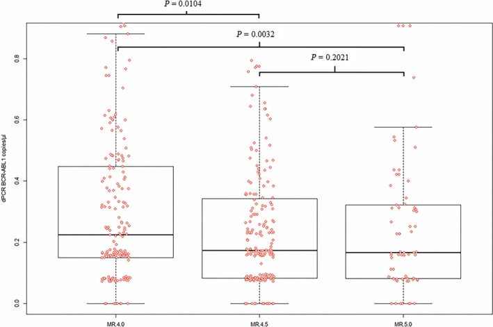
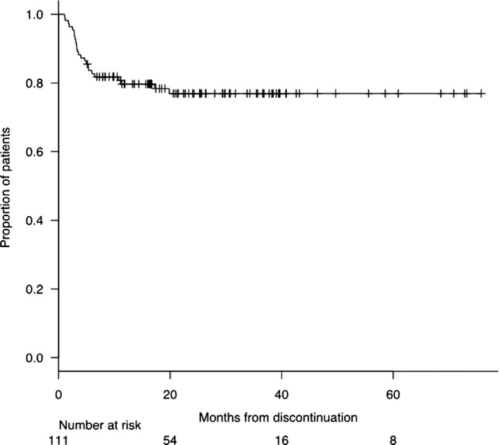
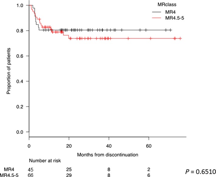
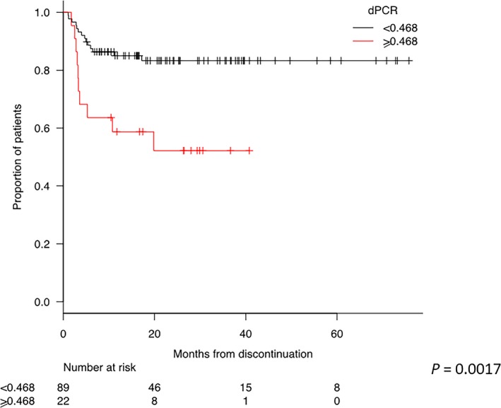
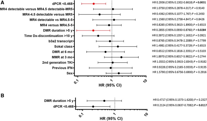
Treatment-free remission (TFR) by tyrosine kinase inhibitors (TKI) discontinuation in patients with deep molecular response (DMR) is a paramount goal in the current chronic myeloid leukemia (CML) therapeutic strategy. The best DMR level by real-time quantitative PCR (RT-qPCR) for TKI discontinuation is still a matter of debate. To compare the accuracy of digital PCR (dPCR) and RT-qPCR for BCR-ABL1 transcript levels detection, 142 CML patients were monitored for a median time of 24 months. Digital PCR detected BCR-ABL1 transcripts in the RT-qPCR undetectable cases. The dPCR analysis of the samples, grouped by the MR classes, revealed a significant difference between MR4.0 and MR4.5 (P = 0.0104) or MR5.0 (P = 0.0032). The clinical and hematological characteristics of the patients grouped according to DMR classes (MR4.0 vs MR4.5-5.0 ) were superimposable. Conversely, patients with dPCR values <0.468 BCR-ABL1 copies/µL (as we previously described) showed a longer DMR duration (P = 0.0220) and mainly belonged to MR4.5-5.0 (P = 0.0442) classes compared to patients with higher dPCR values. Among the 142 patients, 111 (78%) discontinued the TKI treatment; among the 111 patients, 24 (22%) lost the MR3.0 or MR4.0 . RT-qPCR was not able to discriminate patients with higher risk of MR loss after discontinuation (P = 0.8100). On the contrary, according to dPCR, 12/25 (48%) patients with BCR-ABL1 values ≥0.468 and 12/86 (14%) patients with BCR-ABL1 values <0.468 lost DMR in this cohort, respectively (P = 0.0003). Treatment-free remission of patients who discontinued TKI with a dPCR <0.468 was significantly higher compared to patients with dPCR ≥ 0.468 (TFR at 2 years 83% vs 52% P = 0.0017, respectively). In conclusion, dPCR resulted in an improved recognition of stable DMR and of candidates to TKI discontinuation.

Keywords: chronic myeloid leukemia; digital PCR (dPCR); minimal residual disease (MRD) monitoring; treatment-free remission (TFR); tyrosine kinase inhibitors (TKI) discontinuation.


Conflict of interest statement

The authors declare no competing financial interests.

Figures

Figure 1
Figure 1
Levels of BCR‐ABL1 transcript measured by dPCR (y‐axis) according to different MR classes calculated by RT‐qPCR (x‐axis). RT‐qPCR BCR‐ABL1 results were grouped into MR4.0, MR4.5, and MR5.0 class (“X axis”). When the same samples were analyzed by dPCR, different levels of BCR‐ABL1 copies were measured (“Y axis”). The difference was statistically significant between the MR4.0 class and the MR4.5 (P = 0.01) or the MR5.0 class (P = 0.003), but not between the MR4.5 and the MR5.0 classes (P = 0.2). The statistic test used was t test. In box and whiskers plot, center line represents the median values; the boxes’ limits represent the lower and the higher quartile; whiskers define the extreme values; and points represent the single analysis values
Figure 2
Figure 2
Molecular residual disease (MRD) over time as measured by RT‐qPCR and dPCR. (A) MRD monitoring by RT‐qPCR in patients with MR4.0 at Time Point 0. (B) MRD monitoring by RT‐qPCR in patients with MR4.5‐5.0 at Time Point 0. (C) MRD monitoring by dPCR in patients with value of BCR‐ABL1 copies/µL ≥0.468 at Time Point 0. (D) MRD monitoring by dPCR in patients with value of BCR‐ABL1 copies/µL <0.468 at Time Point 0
Figure 3
Figure 3
Treatment‐free remission (TFR) curve of 111 patients in deep molecular response (DMR) who discontinued TKI. The probability of maintaining TFR was 83%, 80%, and 77% at 6, 12, and 24 months, respectively. The plot has been performed by the Kaplan‐Meier method
Figure 4
Figure 4
Treatment‐free remission (TFR) curves according to MR class measured by RT‐qPCR at the time of discontinuation. The red curve represents patients discontinued with MR4.0, and the black curve patients with MR4.5‐5.0. The probability of maintaining TFR for patients discontinued with MR4.0 was 80% at both 1 and 2 years. The probability of maintaining TFR for patients discontinued with MR4.5‐5.0 was 79% and 74% at 1 and 2 years, respectively. Kaplan‐Meier analysis was used for the evaluation of TFR. Comparison of subgroups was carried out by the log‐rank test
Figure 5
Figure 5
Treatment‐free remission (TFR) curves according to dPCR values. The red curve represents patients discontinued with a dPCR value lower than 0.468 and the black curve patients with a dPCR value higher than 0.468. The probability of maintaining TFR for patients discontinued with dPCR <0.468 was 85% and 83% at 1 and 2 years, respectively. The probability of maintaining TFR for patients discontinued with dPCR ≥ 0.468 was 59% and 52% at 1 and 2 years, respectively. A Kaplan‐Meier analysis was used for the evaluation of TFR. Comparison of subgroups was carried out by a log‐rank test
Figure 6
Figure 6
Univariate and multivariate analyses for the prediction of treatment‐free remission (TFR). (A) Univariate Cox regression analysis. Variables included were age at diagnosis, sex, previous therapy with IFN, time to MMR and DMR, Sokal class, type of transcript, time to discontinuation, use of frontline second‐generation TKIs, MR classes considering detectable and undetectable transcript, and dPCR. DMR duration > 5 years (HR 0.2855, CI 95% 0.0931‐0.8760, P = 0.0284) and dPCR (HR 0.2936, CI 95% 0.1302‐0.6618, P = 0.0031) resulted significantly predictable of TFR maintenance. (B) Multivariate analysis—only dPCR retained its significant value (HR 0.2124, CI 95% 0.0637‐0.7082, P = 0.0117)

References

    1. Kantarjian H, O'Brien S, Jabbour E, et al. Improved survival in chronic myeloid leukemia since the introduction of imatinib therapy: a single‐institution historical experience. Blood. 2012;119(9):1981‐1987. - PMC - PubMed
    1. Hehlmann R, Hochhaus A, Baccarani M. European LeukemiaNet. Chronic myeloid leukaemia Lancet. 2007;370(9584):342‐350. - PubMed
    1. Sasaki K, Strom SS, O’Brien S, et al. Relative survival in patients with chronic‐phase chronic myeloid leukaemia in the tyrosine‐kinase inhibitor era: analysis of patient data from six prospective clinical trials. Lancet Haematol. 2015;2(5):e186‐e193. - PMC - PubMed
    1. Baccarani M, Deininger Mw, Rosti G, et al. European LeukemiaNet recommendations for the management of chronic myeloid leukemia: 2013. Blood. 2013;122(6):872‐884. - PMC - PubMed
    1. Latham S, Bartley PA, Budgen B, et al. BCR‐ABL1 expression, RT‐qPCR and treatment decisions in chronic myeloid leukaemia. J Clin Pathol. 2016;69(9):817‐821. - PubMed

Publication types

MeSH terms

Substances