. 2019 Aug 1;40(11):3203-3221.
doi: 10.1002/hbm.24591. Epub 2019 Apr 5.

Altered static and dynamic functional network connectivity in Alzheimer's disease and subcortical ischemic vascular disease: shared and specific brain connectivity abnormalities

Affiliations

Altered static and dynamic functional network connectivity in Alzheimer's disease and subcortical ischemic vascular disease: shared and specific brain connectivity abnormalities

Zening Fu et al. Hum Brain Mapp. .

Abstract

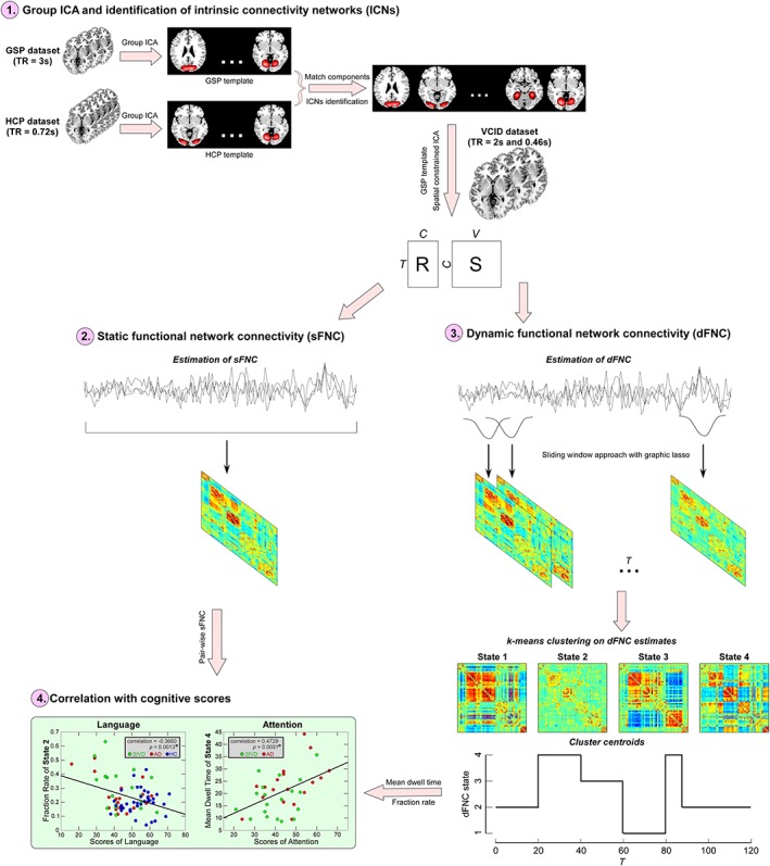
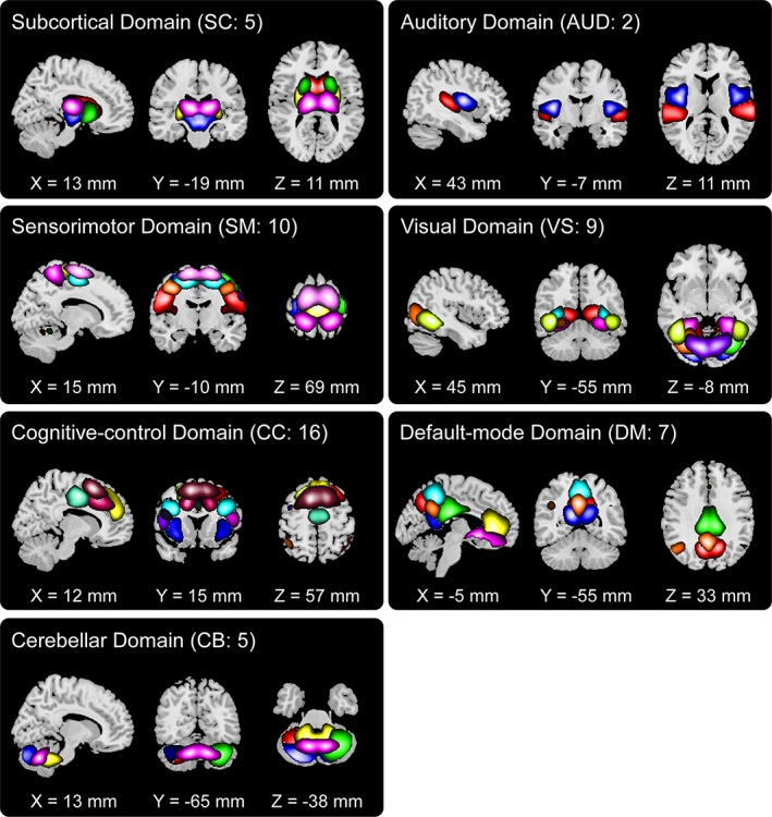
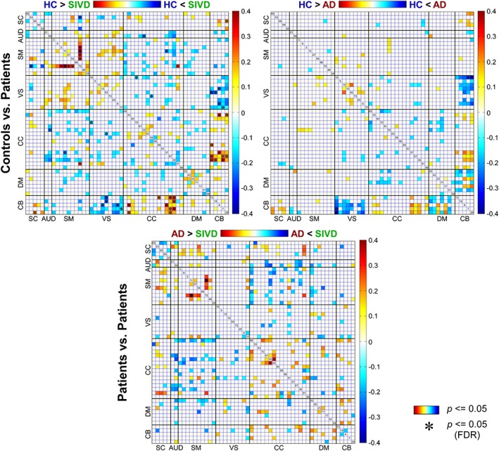
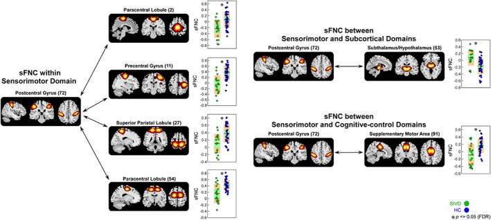
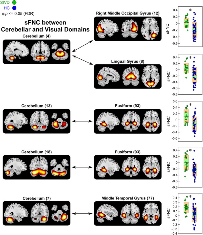
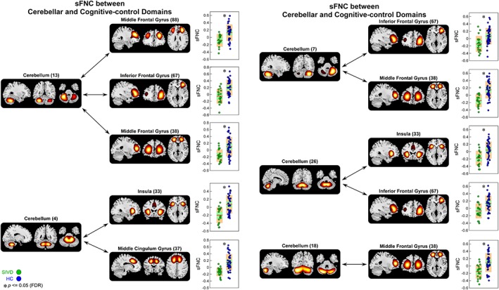
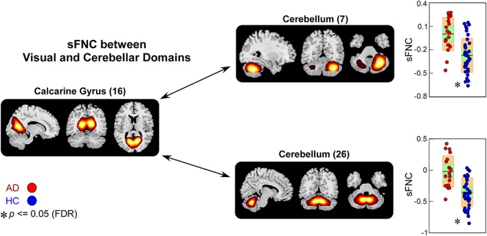
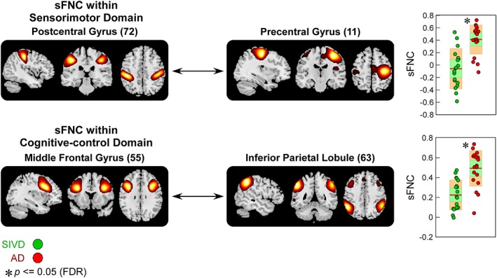
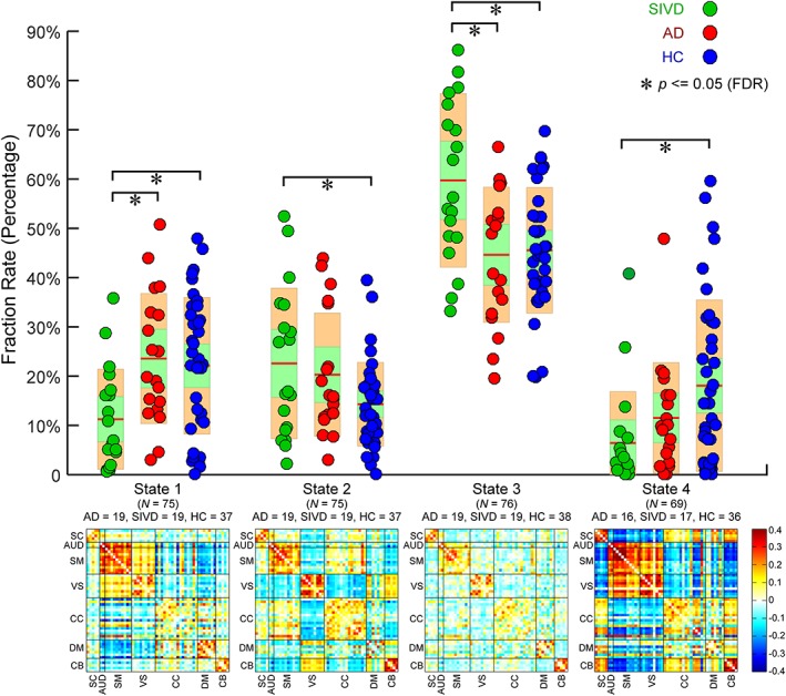
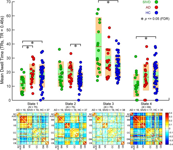
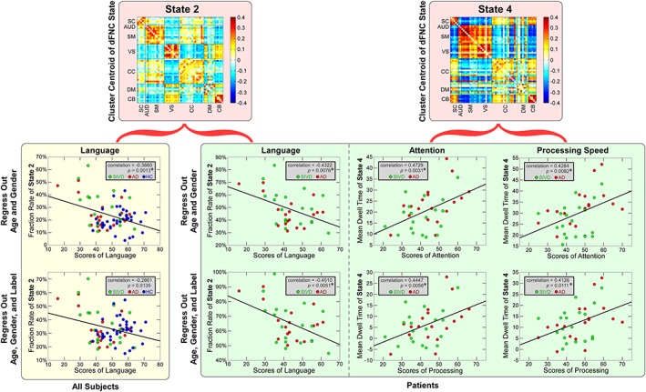
Subcortical ischemic vascular disease (SIVD) is a major subtype of vascular dementia with features that overlap clinically with Alzheimer's disease (AD), confounding diagnosis. Neuroimaging is a more specific and biologically based approach for detecting brain changes and thus may help to distinguish these diseases. There is still a lack of knowledge regarding the shared and specific functional brain abnormalities, especially functional connectivity changes in relation to AD and SIVD. In this study, we investigated both static functional network connectivity (sFNC) and dynamic FNC (dFNC) between 54 intrinsic connectivity networks in 19 AD patients, 19 SIVD patients, and 38 age-matched healthy controls. The results show that both patient groups have increased sFNC between the visual and cerebellar (CB) domains but decreased sFNC between the cognitive-control and CB domains. SIVD has specifically decreased sFNC within the sensorimotor domain while AD has specifically altered sFNC between the default-mode and CB domains. In addition, SIVD has more occurrences and a longer dwell time in the weakly connected dFNC states, but with fewer occurrences and a shorter dwell time in the strongly connected dFNC states. AD has both similar and opposite changes in certain dynamic features. More importantly, the dynamic features are found to be associated with cognitive performance. Our findings highlight similar and distinct functional connectivity alterations in AD and SIVD from both static and dynamic perspectives and indicate dFNC to be a more important biomarker for dementia since its progressively altered patterns can better track cognitive impairment in AD and SIVD.

Keywords: Alzheimer's disease (AD); cognitive impairment; dynamic functional network connectivity (dFNC); resting-state functional network connectivity (rs-FNC); subcortical ischemic vascular disease (SIVD).


Figures

Figure 1
Figure 1
Framework of exploring altered static and dynamic functional network connectivity (FNC) in subcortical ischemic vascular disease (SIVD) and Alzheimer's disease (AD). Step 1: perform group independent component analysis (ICA) on two independent data sets and compute individual spatial maps and time courses using spatially constrained ICA with spatial priors from the exploratory data set; Step 2: estimate static FNC (sFNC) using the Pearson correlation coefficient; Step 3: estimate dynamic FNC (dFNC) using a sliding window approach and perform a k‐means clustering on dFNC estimates; and Step 4: conduct correlation analysis between cognitive scores and sFNC/dFNC features [Color figure can be viewed at http://wileyonlinelibrary.com]
Figure 2
Figure 2
Spatial maps of identified intrinsic connectivity networks (ICNs) are divided into seven different functional domains and arranged based on their anatomical and functional properties. Each color in the composite maps corresponds to a different ICN. The detailed component labels and peak coordinates are provided in Table S1 (see supplementary materials) [Color figure can be viewed at http://wileyonlinelibrary.com]
Figure 3
Figure 3
Group‐discriminating static functional network connectivity (sFNC). Top left: Comparisons between healthy controls (HCs) and patients with subcortical ischemic vascular disease (SIVD); top right: Comparisons between HCs and patients with Alzheimer's disease (AD); bottom: Comparisons between SIVD and AD. sFNC with group difference (p ≤ .05) are highlighted in red and blue. Significant group difference that passes the multiple comparisons is marked by asterisks (false discovery rate [FDR] corrected, q = 0.05) [Color figure can be viewed at http://wileyonlinelibrary.com]
Figure 4
Figure 4
Static functional network connectivity (sFNC) significantly related to healthy control (HC)—subcortical ischemic vascular disease (SIVD) discrimination (regions involved in sensorimotor domain [SM], subcortical domain [SC], and cognitive‐control domain [CC]). Boxplots of sFNC with asterisks indicating significant group difference after false discovery rate (FDR) correction [Color figure can be viewed at http://wileyonlinelibrary.com]
Figure 5
Figure 5
Static functional network connectivity (sFNC) significantly related to healthy control (HC)—subcortical ischemic vascular disease (SIVD) discrimination (regions involved in cerebellar domain [CB] and visual domain [VS]). Boxplots of sFNC with asterisks indicating significant group difference after false discovery rate (FDR) correction [Color figure can be viewed at http://wileyonlinelibrary.com]
Figure 6
Figure 6
Static functional network connectivity (sFNC) significantly related to healthy control (HC)—subcortical ischemic vascular disease (SIVD) discrimination (regions involved in cerebellar domain [CB] and cognitive‐control domain [CC]). Boxplots of sFNC with asterisks indicating significant group difference after false discovery rate (FDR) correction [Color figure can be viewed at http://wileyonlinelibrary.com]
Figure 7
Figure 7
Static functional network connectivity (sFNC) significantly related to healthy control (HC)—Alzheimer's disease (AD) discrimination (regions involved in visual domain [VS] and cerebellar domain [CB]). Boxplots of sFNC with asterisks indicating significant group difference after false discovery rate (FDR) correction [Color figure can be viewed at http://wileyonlinelibrary.com]
Figure 8
Figure 8
Static functional network connectivity (sFNC) significantly related to subcortical ischemic vascular disease (SIVD)—Alzheimer's disease (AD) discrimination (regions involved in sensorimotor domain [SM] and cognitive‐control domain [CC]). Boxplots of sFNC with asterisks indicating significant group difference after false discovery rate (FDR) correction [Color figure can be viewed at http://wileyonlinelibrary.com]
Figure 9
Figure 9
Upper: Group comparisons in fraction rate of occurrences of four dynamic functional network connectivity (dFNC) states. Boxplots of fraction rate of occurrences with asterisks indicating significant group difference after false discovery rate (FDR) correction. Lower: The cluster centroids of four dFNC states, along with the count of subjects that have at least one window clustered into each state [Color figure can be viewed at http://wileyonlinelibrary.com]
Figure 10
Figure 10
Upper: Group comparisons in mean dwell time of four dynamic functional network connectivity (dFNC) states. Boxplots of mean dwell time with asterisks indicating significant group difference after false discovery rate (FDR) correction. Lower: The cluster centroids of four dFNC states, along with the count of subjects that have at least one window clustered into each state [Color figure can be viewed at http://wileyonlinelibrary.com]
Figure 11
Figure 11
The scatterplots illustrate the associations between cognitive scores and each dynamic functional network connectivity (dFNC) feature in the whole samples and in the patient groups. Green circles represent the patients with subcortical ischemic vascular disease (SIVD), red circles represent the patients with Alzheimer's disease (AD), and blue circles represent the healthy controls (HCs). Significant correlations are indicated by asterisks (false discovery rate [FDR] corrected, q = 0.05) [Color figure can be viewed at http://wileyonlinelibrary.com]

References

    1. Agosta, F. , Pievani, M. , Geroldi, C. , Copetti, M. , Frisoni, G. B. , & Filippi, M. (2012). Resting state fMRI in Alzheimer's disease: Beyond the default mode network. Neurobiology of Aging, 33, 1564–1578. 10.1016/j.neurobiolaging.2011.06.007 - DOI - PubMed
    1. Allen, E. A. , Damaraju, E. , Eichele, T. , Wu, L. , & Calhoun, V. D. (2018). EEG signatures of dynamic functional network connectivity states. Brain Topography, 31, 101–116. 10.1007/s10548-017-0546-2 - DOI - PMC - PubMed
    1. Allen, E. A. , Damaraju, E. , Plis, S. M. , Erhardt, E. B. , Eichele, T. , & Calhoun, V. D. (2014). Tracking whole‐brain connectivity dynamics in the resting state. Cerebral Cortex, 24, 663–676. 10.1093/cercor/bhs352 - DOI - PMC - PubMed
    1. Alves, G. S. , Alves, C. E. D. O. , Lanna, M. E. D. O. , Ericeira‐Valente, L. , Sudo, F. K. , Moreira, D. , … Laks, J. (2009). Clinical characteristics in subcortical ischemic white matter disease. Arquivos de Neuro‐Psiquiatria, 67, 173–178. 10.1590/S0004-282X2009000200001 - DOI - PubMed
    1. Anastasi, A. , & Urbina, S. (1997). Psychological testing. Upper Saddle River, NJ, US: Prentice Hall/Pearson Education.

Publication types

MeSH terms