. 2019 Apr 1;75(Pt 4):254-259.
doi: 10.1107/S2053230X1801840X. Epub 2019 Apr 2.

Crystallization of the human tetraspanin protein CD9

Affiliations

Crystallization of the human tetraspanin protein CD9

Rie Umeda et al. Acta Crystallogr F Struct Biol Commun. .

Abstract

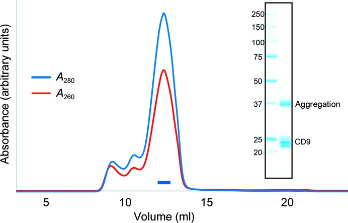
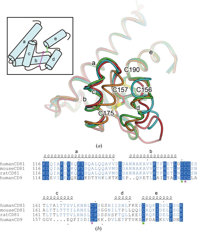
The tetraspanin family of proteins with four membrane-spanning proteins function in a wide range of physiological processes in higher organisms, including cell migration and proliferation, cell fusion, fertilization and virus infection. Although the recently reported structure of CD81 unveiled the basic architecture of this family for the first time, further structural and functional studies are required in order to understand the mechanistic details of the complicated functions of the tetraspanin-family proteins. In this study, attempts were made to crystallize human CD9, a representative member of the tetraspanin family, and it was demonstrated that the truncation of a variable region in the second long extracellular loop significantly improved crystal growth.

Keywords: CD9; LCP; crystallization; membrane proteins; tetraspanins.


Figures

Figure 1
Figure 1
Protein preparation. Size-exclusion chromatogram of wild-type CD9. The blue and red lines indicate the absorbance at 280 and 260 nm, respectively. The blue bar indicates the fractions that were collected and used for crystallization. The inset shows an SDS–PAGE analysis with Coomassie Brilliant Blue staining. Left lane, molecular-mass markers (labeled in kDa); right lane, wild-type CD9. The multiple bands at around 23 kDa represents heterogeneous palmitoylation of the purified CD9 protein, and a higher molecular-weight band at 37 kDa is owing to molecular aggregation during SDS denaturation.
Figure 2
Figure 2
Flexibility of the large extracellular loop. (a) Superimposition of the large extracellular loops of the human CD81 structures [PDB entries 1g8q (green and cyan) and 1iv5 (orange and red)], viewed from the extracellular side. The inset shows a schematic diagram, with green and pink stars representing cysteine residues forming intramolecular disulfide bonds. (b) Amino-acid sequence alignment of the large extracellular loops of human CD81, mouse CD81, rat CD81 and human CD9. Fully and partially conserved residues are highlighted by blue panels and blue letters, respectively. The secondary structure of CD81 is indicated above the alignment. Residues are numbered according to human CD81. Green and pink stars indicate the cysteine residues shown in the diagram in (a).
Figure 3
Figure 3
CD9 crystals and X-ray diffraction. Crystals and X-ray diffraction patterns of native CD9cryst (a) and mercury-derivatized crystals of CD9cryst I20C (b). Scale bars represent 30 µm. The rings indicate 3.5 Å resolution.

References

    1. Caffrey, M. & Cherezov, V. (2009). Nature Protoc. 4, 706–731. - PMC - PubMed
    1. Charrin, S., Jouannet, S., Boucheix, C. & Rubinstein, E. (2014). J. Cell Sci. 127, 3641–3648. - PubMed
    1. Charrin, S., le Naour, F., Silvie, O., Milhiet, P.-E., Boucheix, C. & Rubinstein, E. (2009). Biochem. J. 420, 133–154. - PubMed
    1. Charrin, S., Manié, S., Oualid, M., Billard, M., Boucheix, C. & Rubinstein, E. (2002). FEBS Lett. 516, 139–144. - PubMed
    1. Hemler, M. E. (2003). Annu. Rev. Cell Dev. Biol. 19, 397–422. - PubMed