Review
. 2019 May 6:48:465-494.
doi: 10.1146/annurev-biophys-052118-115534. Epub 2019 Apr 5.

Regulation of Transmembrane Signaling by Phase Separation

Affiliations
Review

Regulation of Transmembrane Signaling by Phase Separation

Lindsay B Case et al. Annu Rev Biophys. .

Abstract

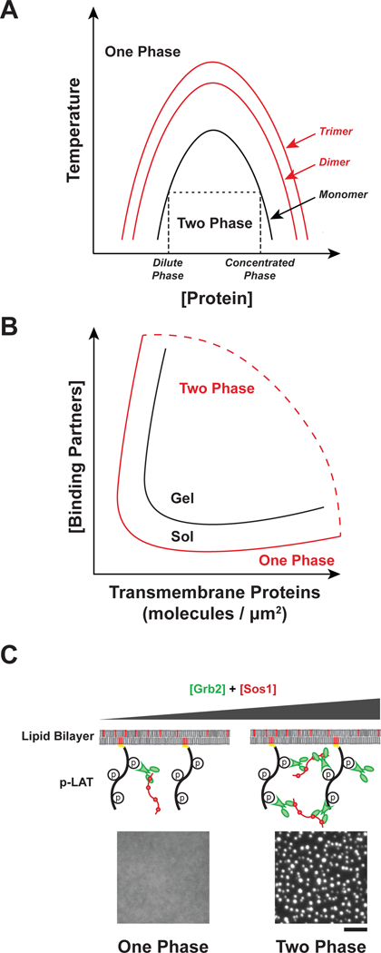
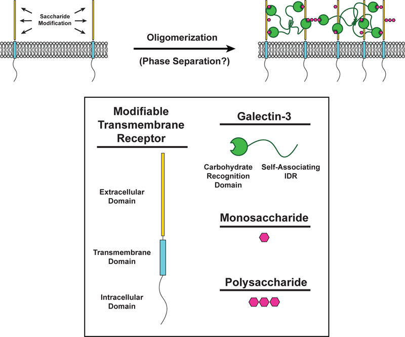
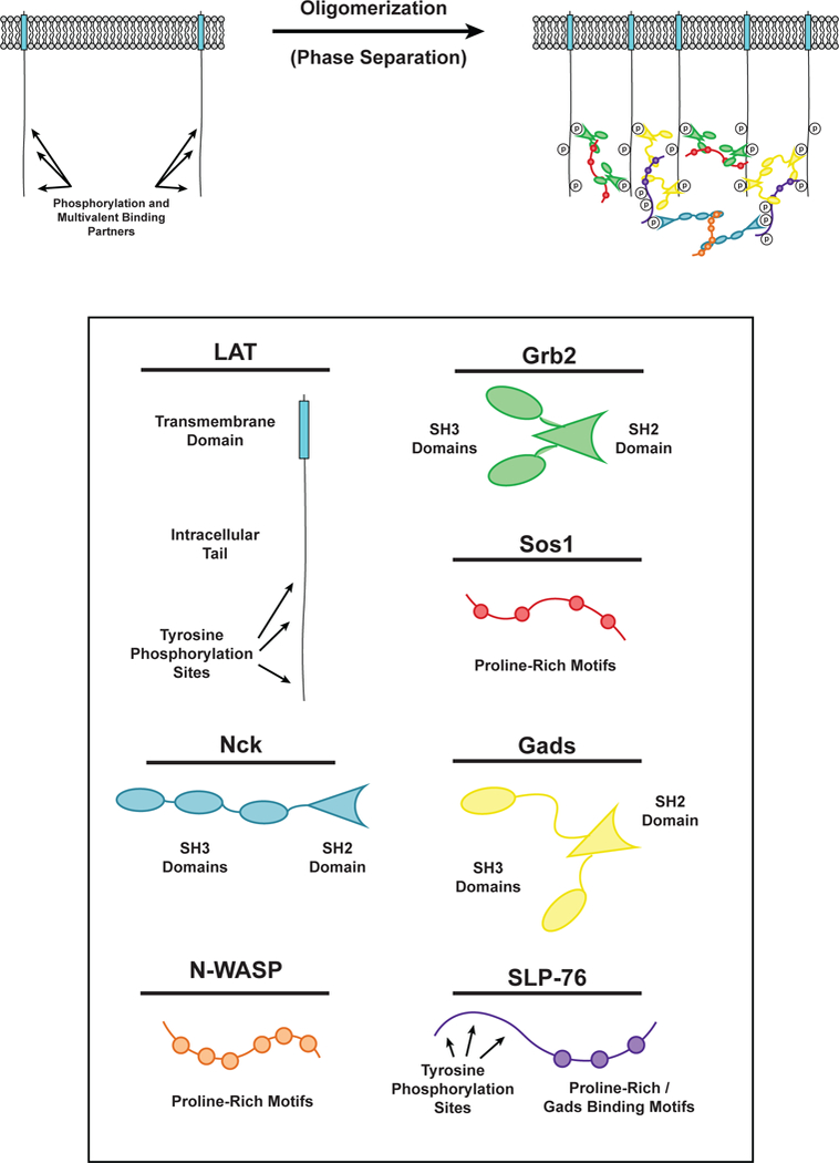
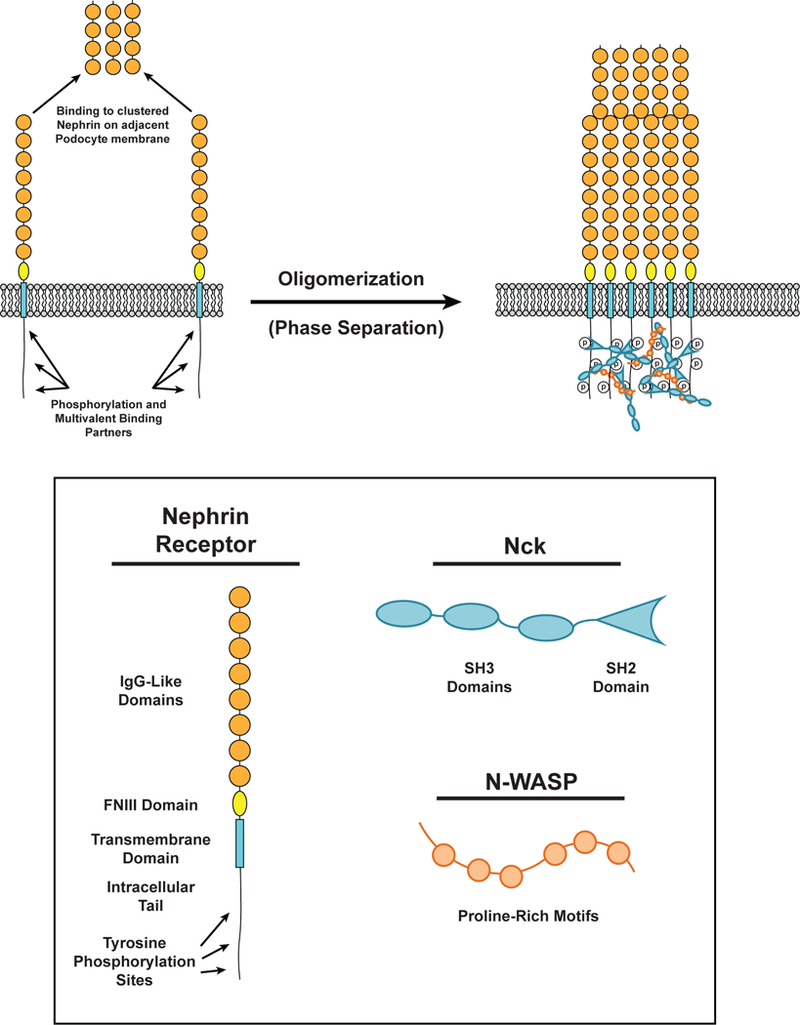
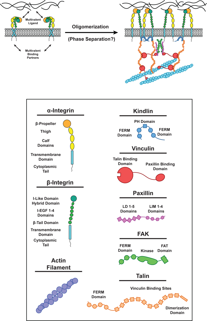
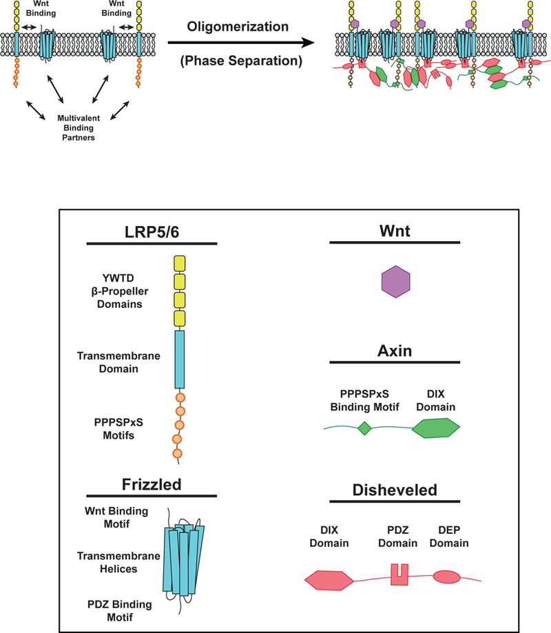
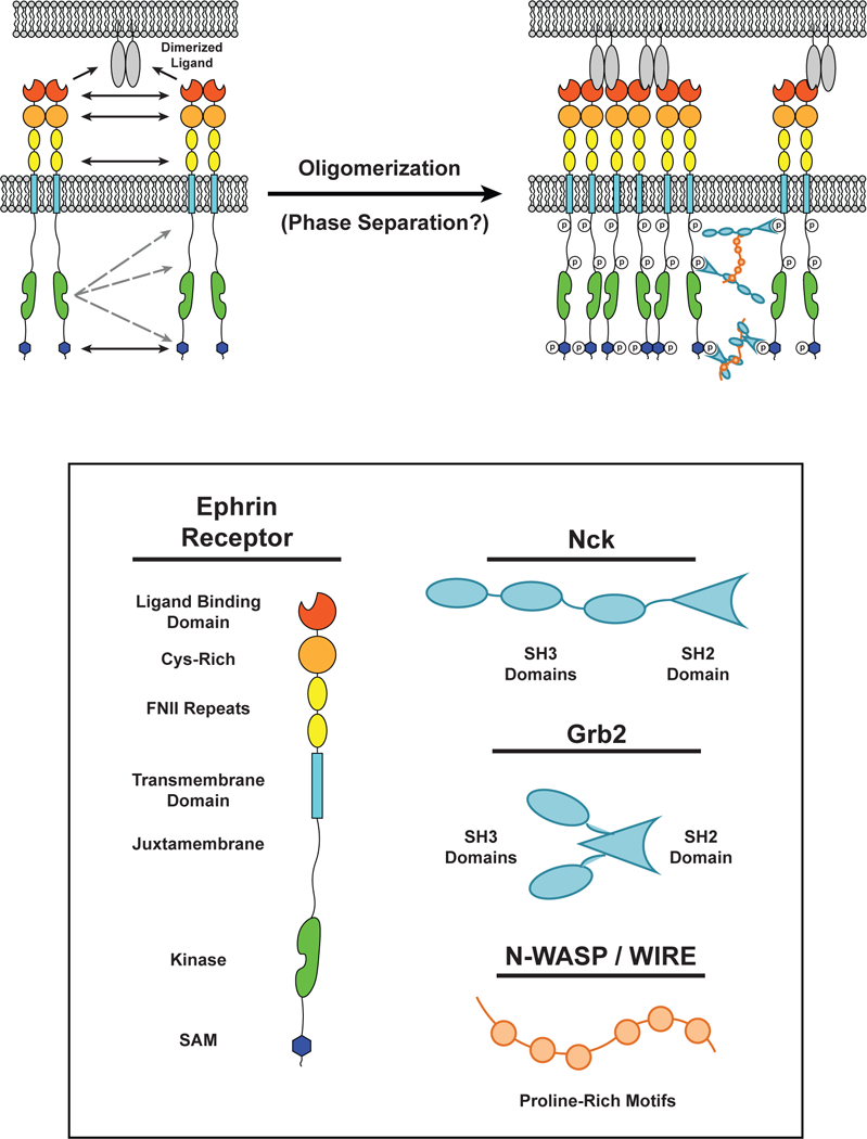
Cell surface transmembrane receptors often form nanometer- to micrometer-scale clusters to initiate signal transduction in response to environmental cues. Extracellular ligand oligomerization, domain-domain interactions, and binding to multivalent proteins all contribute to cluster formation. Here we review the current understanding of mechanisms driving cluster formation in a series of representative receptor systems: glycosylated receptors, immune receptors, cell adhesion receptors, Wnt receptors, and receptor tyrosine kinases. We suggest that these clusters share properties of systems that undergo liquid-liquid phase separation and could be investigated in this light.

Keywords: biomolecular condensates; cell signaling; phase separation; receptor clusters; receptor organization.


Figures

Figure 1.
Figure 1.
Regulation of sol-gel transition and phase separation. A) Oligomerization promotes phase separation. Phase diagram as a function of protein concentration and temperature for monomeric proteins (black line) or oligomers (red lines). Changes to temperature, pH, salt concentration, or crowding can also alter the shape of the binodal curve of the system. At low concentrations, molecules exist in a single dilute phase (One Phase). As concentration increases molecules separate into a dilute phase and a concentrated phase (Two Phase). At high concentrations, molecules exist in a single concentrated phase (One Phase). Phase separation is promoted by increasing the concentration of cytosolic binding partners. B) Phase diagram as a function of transmembrane protein density and cytosolic binding partner concentration. The binodal curve of the sol-gel transition (black) and the phase separation (red) represent distinct physical processes. C) Representative images showing in vitro phase separation of phospho-LAT, Grb2, and Sos1 on membranes. At low Grb2 and Sos1 concentrations the proteins exist in one dilute phase, but at higher concentrations phase separation occurs. Scale bar = 5 μm.
Figure 2.
Figure 2.
Carbohydrate-lectin interactions in receptor oligomerization. (LEFT) The extracellular domain of transmembrane receptors (Yellow) can be modified with monosaccharides and polysaccharides (Magenta) to create binding sites for the carbohydrate recognition domain (CRD) of Galectin-3 (Green). (RIGHT) Saccharide-modified transmembrane receptors are bound by the CRD of Galectin-3. The intrinsically disordered region (IDR) of Galectin-3 self-associates with other IDRs of neighboring Galectin-3 molecules to form a multivalent network of modified transmembrane receptors and Galectin-3.
Figure 3.
Figure 3.
Phase separation of LAT in T cell signaling. (LEFT) LAT transmembrane adaptor proteins can be phosphorylated at three tyrosine residues in the disordered tail to created binding sites for the multivalent adaptor proteins Grb2 (Green) and Gads (Yellow). (RIGHT) Phosphorylated tyrosine residues on LAT can be bound by the SH2 domain of Grb2. The SH3 domains of Grb2 bind to proline-rich motifs of Sos1 (Red) to form a phase-separated clusters of LAT, Grb2, and Sos1. Similarly, phosphorylated tyrosine residues on LAT can be bound by the SH2 domain of Gads. The SH3 domains of Gads bind to proline-rich motifs and a RxxK motif in SLP-76 (Purple) to form a phase-separated cluster of LAT, Gads, and SLP-76. Phosphorylated tyrosine residues on SLP-76 can be bound by the SH2 domain of Nck (Cyan). The SH3 domains of Nck bind to the proline-rich motifs of N-WASP (Orange) to create another level of multivalent interactions that can contribute to the phase separation of LAT on the membrane of T cells.
Figure 4.
Figure 4.
Phase separation in Nephrin in kidney podocytes. (LEFT) The distal-most IgG-like extracellular domain (Orange) of nephrin receptors can bind the distal-most IgG-like extracellular domain of nephrin receptors on the apposing podocyte membrane. The intracellular tail of nephrin is phosphorylated at three tyrosine residues. These phosphorylated residues can be bound by Nck (Cyan). (RIGHT) Phosphorylated tyrosine residues on nephrin can be bound by the SH2 domain of Nck. Nck SH3 domains bind to proline-rich motifs on N-WASP (Orange) to form a phase-separated cluster of nephrin, Nck, and N-WASP. Extracellular interactions between the distal-most IgG-like domains of nephrin also contribute to cluster formation.
Figure 5.
Figure 5.
Multivalent interactions at Focal Adhesions. (LEFT) Integrin receptors in the inactive conformation. (RIGHT) Integrin receptors can be activated when the extracellular domain binds multivalent components of the extracellular matrix (ECM) and the intraceullar domain of β-integrin binds Kindlin (Blue) and/or Talin (Orange). Kindlin dimerizes by interactions in the FERM domain. Talin is composed of ten Vinculin (Red) binding sites and can bind actin filaments by a site in its C-terminus dimerization domain. Vinculin can bind Talin (via its N-terminal Talin binding domain) and Paxillin (Purple) LD motifs and actin filaments by a site on its C-terminus. Paxillin LD motifs can bind the Vinculin C-terminus and FAK FAT domains. FAK (Green) can dimerize by interactions between its FERM domains. Each of these unique interactions between multiple proteins results in the formation of a highly interconnected oligomeric protein network in focal adhesions.
Figure 6.
Figure 6.
Phase separation in Wnt Signaling. (LEFT) Wnt (Purple) can bind to LRP5/6 (single-pass transmembrane receptor) and Frizzled (Fz) receptors (multipass transmembrane receptor) to initiate Wnt signaling. LRP5/6 can bind Axin (Green) while Fz can bind Disheveled (Dvl, Red). These interactions induce the formation of phase-separated clusters on the cell membrane. (RIGHT) Following Wnt binding, phosphorylation of PPPSPxS motifs on LRP5/6 enables binding of Axin via an undefined region of its C-terminus. Disheveled can bind the Fz receptor intracellular tail by its PDZ domain. Polymerization of DIX domains in both Axin and Dvl promote clustering of membrane-associated proteins at Fz and LRP5/6 receptors. Dvl also contains a C-terminal dimerization domain that enhances multivalent interactions within the Wnt signalosome.
Figure 7.
Figure 7.
Multiple interactions drive dimerization and cluster formation of ephrin receptors. (LEFT) Interactions between two ephrin receptor EC ligand binding domains (LBDs), Cys-rich domains, and fibronectin-like (FN) domains can result in ephrin receptor dimerization in the absence of ligands. Dimerized ligands expressed on the surface of an apposing cell can seed cluster formation. (RIGHT) Upon binding to a ligand, the kinase domain of one ephrin receptor can transphosphorylate intracellular tyrosine residues on surrounding receptors. These residues can serve as docking sites for Src Homolgy (SH) SH2 / SH3 adaptor proteins, such as Grb2 and Nck, which can oligomerize through multivalent interactions with proteins containing Proline-Rich Motifs (PRMs), such as N-WASP. The intracellular Sterile Alpha Motifs (SAMs) also self-associate to promote receptor oligomerization.

References

    1. Ahmad N, Gabius H-J, André S, Kaltner H, Sabesan S, et al. 2004. Galectin-3 Precipitates as a Pentamer with Synthetic Multivalent Carbohydrates and Forms Heterogeneous Cross-linked Complexes. J. Biol. Chem. 279(12):10841–47 - PubMed
    1. Asherie N, Pande J, Lomakin A, Ogun O, Hanson SRA, et al. 1998. Oligomerization and phase separation in globular protein solutions. Biophys. Chem. 75(3):213–27 - PubMed
    1. Atanasova M, Whitty A. 2012. Understanding cytokine and growth factor receptor activation mechanisms. Crit. Rev. Biochem. Mol. Biol. 47(6):502–30 - PMC - PubMed
    1. Balagopalan L, Kortum RL, Coussens NP, Barr VA, Samelson LE. 2015. The Linker for Activation of T Cells (LAT) Signaling Hub: From Signaling Complexes to Microclusters. J. Biol. Chem. 290(44):26422–29 - PMC - PubMed
    1. Banani SF, Lee HO, Hyman AA, Rosen MK. 2017. Biomolecular condensates: organizers of cellular biochemistry. Nat. Rev. Mol. Cell Biol. 18(5):285–98 - PMC - PubMed

Publication types

LinkOut - more resources