. 2019 May 21;50(5):1249-1261.e5.
doi: 10.1016/j.immuni.2019.03.002. Epub 2019 Apr 2.

Keratinocyte-Mediated Activation of the Cytokine TGF-β Maintains Skin Recirculating Memory CD8+ T Cells

Affiliations

Keratinocyte-Mediated Activation of the Cytokine TGF-β Maintains Skin Recirculating Memory CD8+ T Cells

Toshiro Hirai et al. Immunity. .

Abstract

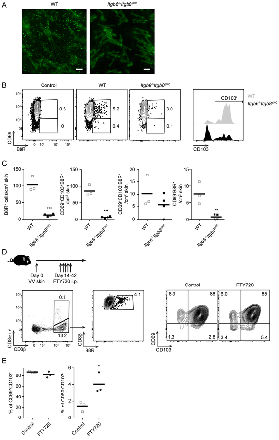
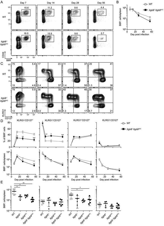
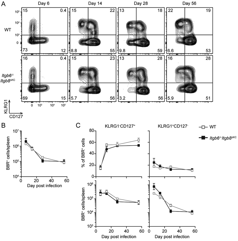
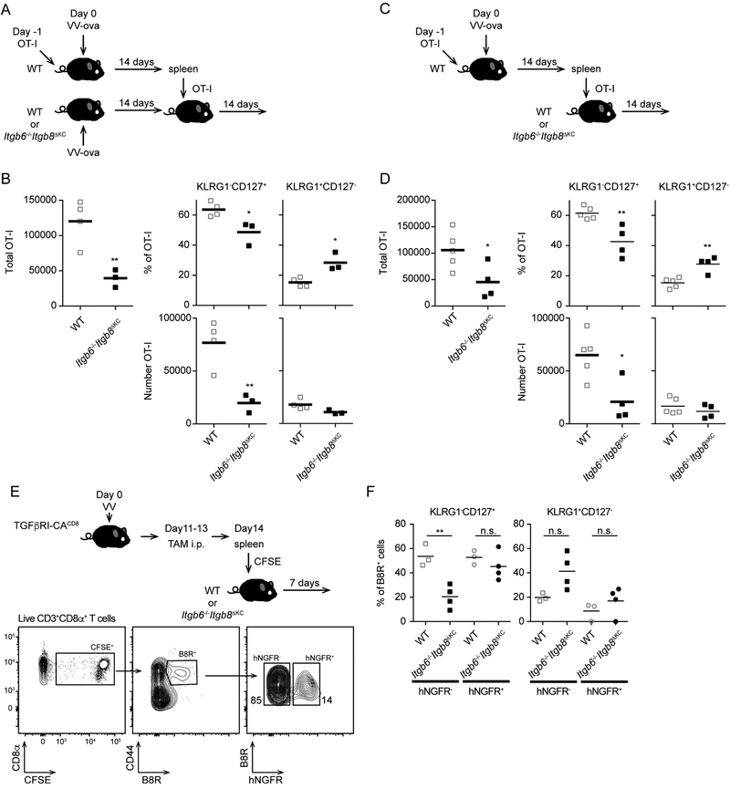
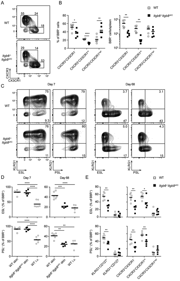
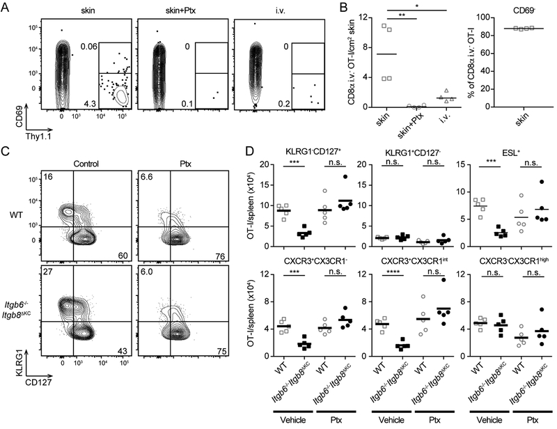
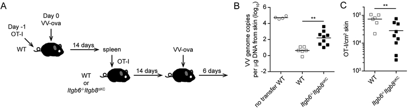
Regulated activation of the cytokine TGF-β by integrins αvβ6 and αvβ8 expressed on keratinocytes is required for residence of epidermal-resident memory T cells, but whether skin-derived signals also affect recirculating memory cells in the skin remains unclear. Here, we show that after resolution of skin vaccinia virus (VV) infection, antigen-specific circulating memory CD8+ T cells migrated into skin. In mice lacking αvβ6 and αvβ8 integrins (Itgb6-/-Itgb8fl/fl-K14-cre), the absence of epidermal-activated TGF-β resulted in a gradual loss of E- or P-selectin-binding central and peripheral memory populations, which were rescued when skin entry was inhibited. Skin recirculating memory cells were required for optimal host defense against skin VV infection. These data demonstrate that skin migration can persist after resolution of local skin infection and that the cytokine environment within this nonlymphoid tissue shapes the differentiation state and persistence of the central and peripheral memory-T-cell pool.

Keywords: CD8(+) T cell memory; keratinocytes; skin; transforming growth factor beta; α(v)β(6); α(v)β(8).


Conflict of interest statement

Declaration of Interests

The authors declare no competing interests.

Figures

Figure 1.
Figure 1.. Trm cells and recirculating CD8+ T cells are decreased in the skin of Itgb6−/−Itgb8ΔKC mice after skin vaccinia virus (VV)-infection
(A) Representative epidermal whole mounts from OT-I adoptive transferred WT and Itgb6−/−Itgb8ΔKC mice at day 42 after VV-OVA skin infection stained for OT-I cells (Thy1.1, green). Scale bar=100 μm. (B) VV-infected skin at day 68 post infection were analyzed by flow cytometry. Plots (left panels) are gated on live CD45+CD3+TCRb+CD8+ T cells (WT and Itgb6−/−Itgb8ΔKC) or CD45+CD3+TCRb+CD8 T cells (control), and B8R+ CD69+ (right panel). (C) Total number of B8R+ cells from (B) are shown. (D) Experimental scheme for testing skin recirculating CD8+ T cells in VV-infected skin. Anti-CD8α antibody was intravenously injected to stain intravascular CD8+ T cells before harvesting VV-infected skin for flow cytometric analysis one day after the last treatment of FTY720. Flow plots gated on live CD90+ T cells (left panel), i.v. CD8αCD8β+ (middle panel), and B8R+ cells (right panels). (E) The frequency of i.v. CD8αB8R+ gated cells in the skin are shown. Data are representative of two independent experiments. Each dot represents an individual mouse. Flow cytometry plots are shown as concatenations for all mice in each group within the same experiment. *p < 0.05, **p < 0.01, and ***p < 0.001. See also Figure S1.
Figure 2.
Figure 2.. Vaccinia specific splenic CD8+ T cells are not maintained in Itgb6−/−Itgb8ΔKC mice
Spleens from WT or Itgβ6−/−Itgβ8ΔKC mice were analyzed by flow cytometry at the indicated time point after skin VV infection. Representative plots gated on live CD3+CD8+ cells (A) the total number of B8R+ cells (B) at each time point are shown. (C) Representative plots gated on B8R+ cells from (A) are shown. (D) The frequency (upper panels) and number (lower panels) of splenic B8R+ cells are shown. (E) Splenic B8R+ cells from WT, Itgβ6−/−, Itgβ8ΔKC, or Itgβ6−/−Itgβ8ΔKC mice were analyzed by flow cytometry at 68 days after skin VV infection. Data are representative of two or three independent experiments with at least four mice per time point. Data are means ± s.e.m. *p < 0.05, **p < 0.01, and ***p < 0.001. See also Figure S2.
Figure 3.
Figure 3.. Intravenous vaccinia virus (VV)-induced CD8+ T cells are maintained normally in Itgb6−/−Itgb8ΔKC mice
Splenic cells at the indicated time following intravenous VV infection in WT or Itgb6−/−Itgb8ΔKC mice were analyzed for KLRG1CD127+ memory precursors and KLRG1+CD127 terminal effectors by flow cytometry. (A) Representative plots gated on B8R+ cells are shown. (B) Total number of B8R+ cells, and (C) frequency (top panels) and total number (bottom panels) of KLRG1CD127+ or KLRG1+CD127 B8R+ cells are shown. Data are representative of two separate experiments with at least four mice per each time point. Data are means ± s.e.m.
Figure 4.
Figure 4.. KLRG1CD127+ Memory precursors are not maintained following adoptive transfer into integrin-deficient mice.
(A) The experimental scheme for adoptive transfer into matched infected is shown. OT-I adoptive transfer WT mice were infected with VV-OVA. On day 14 post infection, splenic CD8+ T cells were isolated and CD8+ T cells containing 2 × 10⁶ OT-I cells were adoptively transferred into matched infected WT or Itgb6−/−Itgb8ΔKC mice. (B) Total numbers of Thy1.1+OT-I cells (left panel) from the spleens of recipient WT or Itgb6−/−Itgb8ΔKC mice 28 days after infection (14 days post transfer) are shown (left panel). The percentage (top panels) and total number (bottom panels) of KLRG1+CD127 and KLRG1CD127+ OT-I cells are shown. (C) The experimental scheme for adoptive transfer into naive mice is shown. As in (A) except cells were adoptively transferred into naïve WT or Itgb6−/−Itgb8ΔKC recipients. (D) Total numbers of Thy1.1+OT-I cells from the spleens of recipient WT or Itgb6−/−Itgb8ΔKC mice 28 days after infection (14 days post transfer) is shown (left panel). The percentage (top, right panels) and total number (bottom, right panels) of KLRG1+CD127 and KLRG1CD127+ OT-I cells is shown. (E) Experimental scheme for testing a direct requirement for TGFβ on skin recirculating memory cells. Flow plots shows gating strategy for analysis of the transferred CFSE+B8R+ cells expressing TGFβRI-CA (hNGFR+) or control (hNGFR) in spleen day 7 post transfer. (F) Frequency of the indicated subsets of CFSE+B8R+ cells are shown. Data are representative of two to four independent experiments. Each dot represents an individual mouse. *p < 0.05 and **p < 0.01. See also Figure S3, S4, and S5.
Figure 5.
Figure 5.. Skin-homing memory cells are decreased in Itgb6−/−Itgb8ΔKC mice
(A and B) Splenic B8R+ cells from WT or Itgb6−/−Itgb8ΔKC mice were analyzed for memory subsets based on expression of CXCR3 and CX3XR1 by flow cytometry at day 68 post skin vaccinia virus (VV) infection: CXCR3+CX3CR1 Tcm cells, CXCR3+CX3CR1int Tpm cells and CXCR3CX3CR1high Tem cells. (A) Representative flow cytometry plots gated on B8R+ cells and the (B, left panel) frequency and (B, right panel) number of each memory subset of B8R+ cells are shown. (C and D) E-selectin or P-selectin binding (ESL+ or PSL+ respectively) of splenic B8R+ cells were analyzed by flow cytometry following skin VV infection. Representative flow plots (C) and the frequency of ESL+ (top panels) or PSL+ (bottom panels) B8R+ cells day 7 and 68 post infection are shown. (D) Frequency of ESL+ (top panels) or PSL+ (bottom panels) among each subset of memory B8R+ cells in the spleen are shown. Data are representative of two separate experiments. Each dot represents an individual mouse. *p < 0.05, **p < 0.01, ***p < 0.001, and ****p < 0.0001. See also Figure S6.
Figure 6.
Figure 6.. Skin recirculating CD8+ T cells require TGF-β activation by keratinocytes for maintenance
(A and B) Naïve Thy1.1+OT-I cells were transferred to WT mice followed by skin or intravenous vaccinia virus (VV)-OVA infection. Total CD8+ T cells were collected from spleens 14 days post VV-OVA infection and 106 OT-I cells treated with vehicle or pertussis toxin (Ptx) (100 ng/mL) were adoptively transferred to naïve WT recipient mice. Anti-CD8α antibody was intravenously injected to stain intravascular CD8+ T cells before harvesting skin for flow cytometric analysis. (A) Representative plot of OT-I cells from the skin of recipient mice 7 days post transfer gated on live i.v. CD8αCD8β+ cells are shown. (B) The total number of OT-I cells and frequency of CD69 OT-I cells in the skin is shown. (C and D) Naïve Thy1.1+OT-I cells were transferred to WT mice followed by skin VV-OVA infection. Total CD8+ T cells were isolated from spleens 14 days post skin VVOVA infection and treated with vehicle or pertussis toxin (Ptx) (100 ng/mL) immediately before adoptive transfer (106 OT-I/mouse) into naïve WT or Itgb6−/−Itgb8ΔKC recipients. (C) Representative plots and (D) total numbers of OT-I cells in the spleen 7 days after transfer are shown. Data are representative of two or three separate experiments. Each dot represents an individual recipient. n.s.; not significant, *p < 0.05, **p < 0.01, ***p < 0.001, and ****p < 0.0001. See also Figure S7.
Figure 7.
Figure 7.. Skin-recirculating memory CD8+ T cells contribute to host defense against skin VV infection
(A) The experimental scheme is shown. OT-I adoptive transfer WT mice were infected with skin vaccinia virus (VV)-OVA. On day 14 post infection, splenic CD8+ T cells were isolated and CD8+ T cells containing 106 OT-I cells were adoptively transferred into naive WT or Itgb6−/−Itgb8ΔKC recipient mice. Fourteen days after the transfer, the mice were challenged with skin VV-OVA and the infected skin were harvested 6 days after the challenge. (B) qPCR for VV DNA at the infected skin site of WT and Itgb6−/−Itgb8ΔKC mice is shown. (C) Total numbers of OT-I cells identified by flow cytometry in the skin are shown. Data are representative of two separate experiments. Each symbol represents an individual recipient. **p < 0.01.

References

    1. Aluwihare P, Mu Z, Zhao Z, Yu D, Weinreb PH, Horan GS, Violette SM, Munger JS, 2009. Mice that lack activity of alphavbeta6- and alphavbeta8-integrins reproduce the abnormalities of Tgfb1- and Tgfb3-null mice. J. Cell. Sci 122, 227–232. doi:10.1242/jcs.035246 - DOI - PMC - PubMed
    1. Anderson KG, Mayer-Barber K, Sung H, Beura L, James BR, Taylor JJ, Qunaj L, Griffith TS, Vezys V, Barber DL, Masopust D, 2014. Intravascular staining for discrimination of vascular and tissue leukocytes. Nature Protocols 9, 209–222. doi:10.1038/nprot.2014.005 - DOI - PMC - PubMed
    1. Bartholin L, Cyprian FS, Vincent D, Garcia CN, Martel S, Horvat B, Berthet C, Léon SG, Treilleux I, Rimokh R, Marie JC, 2008. Generation of mice with conditionally activated transforming growth factor beta signaling through the TβRI/ALK5 receptor. genesis 46, 724–731. doi:10.1002/dvg.20425 - DOI - PubMed
    1. Bromley SK, Thomas SY, Luster AD, 2005. Chemokine receptor CCR7 guides T cell exit from peripheral tissues and entry into afferent lymphatics. Nature Immunology 6, 895–901. doi:10.1038/ni1240 - DOI - PubMed
    1. Bromley SK, Yan S, Tomura M, Kanagawa O, Luster AD, 2013. Recirculating memory T cells are a unique subset of CD4+ T cells with a distinct phenotype and migratory pattern. J Immunol 190, 970–976. doi:10.4049/jimmunol.1202805 - DOI - PMC - PubMed

Publication types

MeSH terms