Review
. 2019 Sep-Oct;13(5):281-287.
doi: 10.1016/j.jcct.2019.03.007. Epub 2019 Mar 29.

Coronary atherosclerosis imaging by CT to improve clinical outcomes

Affiliations
Review

Coronary atherosclerosis imaging by CT to improve clinical outcomes

Michelle C Williams et al. J Cardiovasc Comput Tomogr. 2019 Sep-Oct.

Abstract

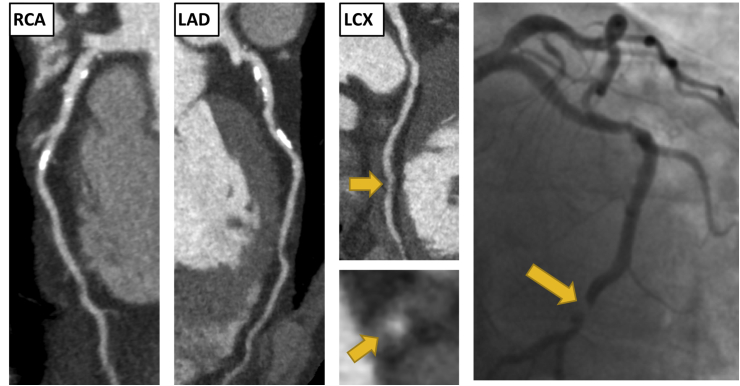
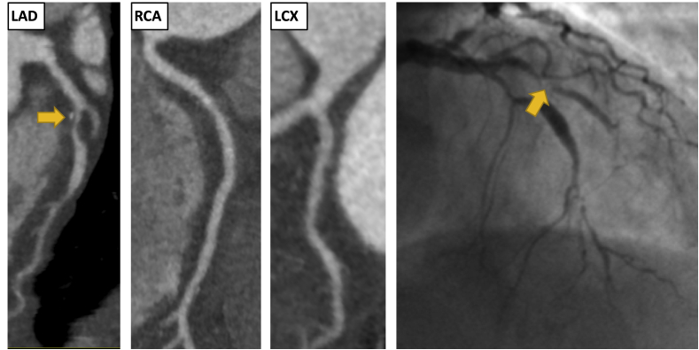
Coronary artery disease remains an important cause of morbidity and mortality world-wide. Coronary Computed Tomography Angiography (CCTA) has excellent diagnostic accuracy and the identification and stratification of coronary artery disease is associated with improved prognosis in multiple studies. Recent randomized controlled trials have shown that in patients with stable coronary artery disease, CCTA is associated with improved diagnosis, changes in investigations, changes in medical treatment and appropriate selection for revascularization. Importantly this diagnostic approach reduces the long-term risk of fatal and non-fatal myocardial infarction. The identification of adverse plaques on CCTA is known to be associated with an increased risk of acute coronary syndrome, but does not appear to be predictive of long-term outcomes independent of coronary artery calcium burden. Future research will involve the assessment of outcomes after CCTA in patients with acute chest pain and asymptomatic patients. In addition, more advanced quantification of plaque subtypes, vascular inflammation and coronary flow dynamics may identify further patients at increased risk.


Figures

Fig. 1
Fig. 1
Clinical Vignette 1. A 65-year-old male presented to the rapid access chest pain clinic with symptoms of typical angina. He was a current smoker with a past medical history of gout. An exercise tolerance test was stopped early due to knee pain, but demonstrated no other abnormalities. His coronary artery calcium score was 283 Agatston units. He underwent a CCTA which showed moderate non-obstructive disease (CAD-RADS 3) He had mild calcified plaque in the left anterior descending (LAD) and right coronary artery (RCA), <50% stenosis. He had moderate non-calcified plaque with positive remodelling in the proximal and distal left circumflex artery (LCX), 50–70% stenosis. He presented 1 years later with an episode of acute chest pain on the background of 2 weeks of worsening chest pain. Electrocardiogram (ECG) on admission showed ST depression in the inferior leads. High sensitivity troponin I was elevated at 2060 ng/L. He was diagnosed with a non ST elevation myocardial infarction (NSTEMI). Invasive coronary angiography showed an occluded mid LCX (yellow arrows). There was also mild disease in the LAD and moderate disease in the small RCA. A bare metal stent was inserted into the LCX and he was asymptomatic on subsequent follow-up. . (For interpretation of the references to colour in this figure legend, the reader is referred to the Web version of this article.)
Fig. 2
Fig. 2
Clinical Vignette 2. A 67-year-old female presented to the rapid access chest pain clinic with atypical chest pain. She had hypertension but no other cardiovascular risk factors. Her estimated 10-year risk of cardiovascular disease was 14% (ASSIGN score). An exercise tolerance test was normal. Her coronary artery calcium score was 62 Agatston units. CCTA identified single vessel obstructive coronary artery disease (CAD-RADS 4A). There was severe mixed plaque in the proximal and mid LAD, >70% stenosis. The plaque in the proximal LAD demonstrated positive remodelling and spotty calcification. There was also mild disease in the mid RCA and proximal LCX, <50% stenosis. 4 years later she presented to the emergency department with acute chest pain. ECG showed ST depression in the inferior leads. High sensitivity troponin I was elevated at 387 ng/L. Invasive coronary angiogram showed an occluded LAD (yellow arrows) and mild disease in the LCX and RCA. The LAD was treated with a drug eluting stent. She had no further hospital admissions in the subsequent 3 years. . (For interpretation of the references to colour in this figure legend, the reader is referred to the Web version of this article.)

References

    1. WHO. World Health Statistics . Sustainable Development Goals; 2018. Monitoring Health for the SDGs; pp. 1–9. 2018.
    1. Miller J.M., Rochitte C.E., Dewey M. Diagnostic performance of coronary angiography by 64-row CT. N Engl J Med. 2008;359(22):2324–2336. - PubMed
    1. Dewey M., Rief M., Martus P. Evaluation of computed tomography in patients with atypical angina or chest pain clinically referred for invasive coronary angiography: randomised controlled trial. BMJ. 2016;355:i5441. - PMC - PubMed
    1. Andreini D., Pontone G., Mushtaq S. A long-term prognostic value of coronary CT angiography in suspected coronary artery disease. JACC (J Am Coll Cardiol): Cardiovascular Imaging. 2012;5(7):690–701. - PubMed
    1. Min J.K., Shaw L.J., Devereux R.B. Prognostic value of multidetector coronary computed tomographic angiography for prediction of all-cause mortality. J Am Coll Cardiol. 2007;50(12):1161–1170. - PubMed

MeSH terms