. 2019 Jun 6;133(23):2529-2541.
doi: 10.1182/blood.2019000424. Epub 2019 Apr 5.

Red blood cells modulate structure and dynamics of venous clot formation in sickle cell disease

Affiliations

Red blood cells modulate structure and dynamics of venous clot formation in sickle cell disease

Camille Faes et al. Blood. .

Abstract

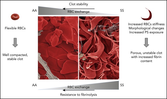
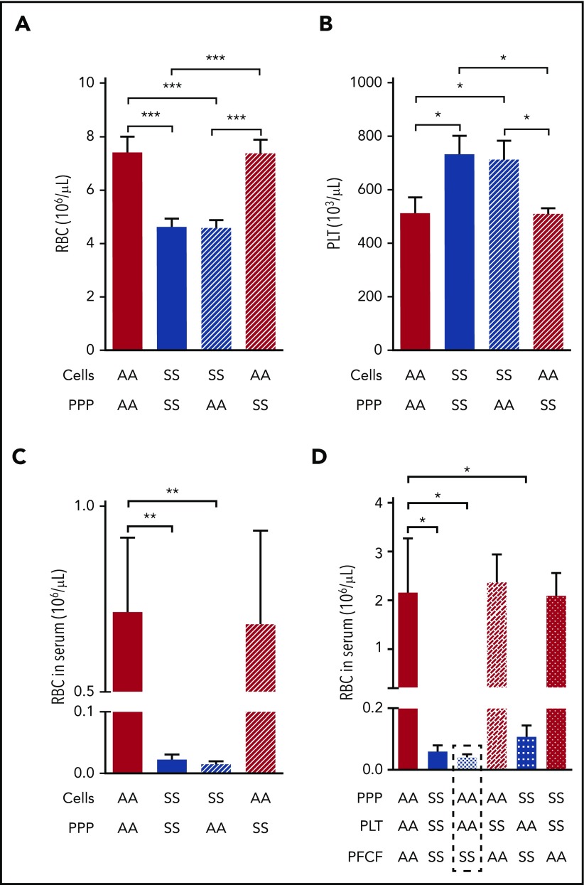
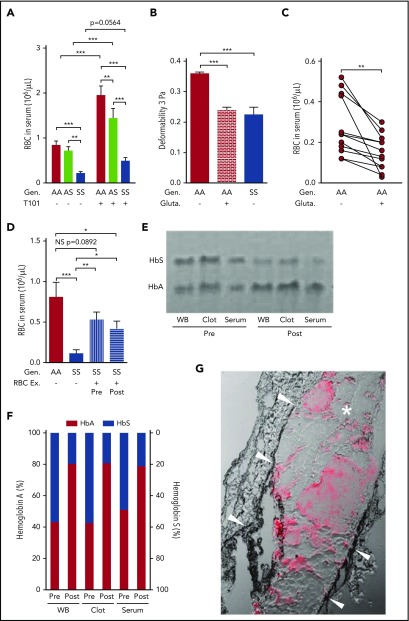
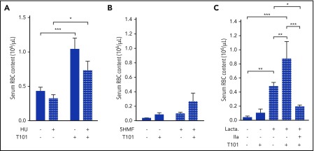
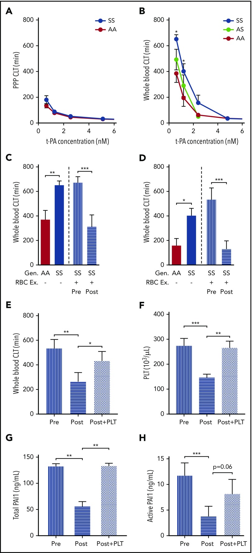
Sickle cell disease (SCD) is associated with chronic activation of coagulation and an increased risk of venous thromboembolism. Erythrocyte sickling, the primary pathologic event in SCD, results in dramatic morphological changes in red blood cells (RBCs) because of polymerization of the abnormal hemoglobin. We used a mouse model of SCD and blood samples from sickle patients to determine if these changes affect the structure, properties, and dynamics of sickle clot formation. Sickling of RBCs and a significant increase in fibrin deposition were observed in venous thrombi formed in sickle mice. During ex vivo clot contraction, the number of RBCs extruded from sickle whole blood clots was significantly reduced compared with the number released from sickle cell trait and nonsickle clots in both mice and humans. Entrapment of sickled RBCs was largely factor XIIIa-independent and entirely mediated by the platelet-free cellular fraction of sickle blood. Inhibition of phosphatidylserine, but not administration of antisickling compounds, increased the number of RBCs released from sickle clots. Interestingly, whole blood, but not plasma clots from SCD patients, was more resistant to fibrinolysis, indicating that the cellular fraction of blood mediates resistance to tissue plasminogen activator. Sickle trait whole blood clots demonstrated an intermediate phenotype in response to tissue plasminogen activator. RBC exchange in SCD patients had a long-lasting effect on normalizing whole blood clot contraction. Furthermore, RBC exchange transiently reversed resistance of whole blood sickle clots to fibrinolysis, in part by decreasing platelet-derived PAI-1. These properties of sickle clots may explain the increased risk of venous thromboembolism observed in SCD.


Conflict of interest statement

Conflict-of-interest disclosure: The authors declare no competing financial interests.

Figures

None
Graphical abstract
Figure 1.
Figure 1.
Sickling of RBC and increased deposition of acellular material within venous thrombi formed in SCD mice. Scanning electron microscopy of clots formed 2 hours after inferior vena cava stenosis in AA (A) and SS mice (B-D). Original magnification: ×8000 (A), ×5000 (B-C), and ×20 000 (D).
Figure 2.
Figure 2.
Increased fibrin deposition and evidence of RBC sickling in thrombi formed in the femoral vein of SCD mice. (A) Representative images of platelet (green) and fibrin (red) deposition within thrombi formed in the femoral vein of AA, AS, and SS mice 60 minutes after electrolytic injury. Yellow color shows overlap between fibrin and platelets. Representative movies demonstrating the dynamics of clot formation over 1 hour are included as supplemental Videos 2-4 for AA, AS, and SS mice, respectively. Intravital microscopy analysis of platelet accumulation (B) and fibrin deposition (C) within thrombi of AA (n = 14, gray line), AS (n = 9, purple line), and SS (n = 10, blue line) mice quantitated as a relative intensity. *P < .05. (D) Hematoxylin and eosin staining of the thrombus (delineated by black dotted line) formed in the femoral vein of SS mice demonstrates the presence of RBCs (dark pink) and fibrin/platelets (light pink). Extensive sickling of the RBCs is present within the thrombus (E) but not in the blood pool outside the thrombus (F). Original magnification: ×4 (D) and ×40 (E-F). NS, not significant.
Figure 3.
Figure 3.
Inhibition of clot contraction-mediated extrusion of mouse sickle RBCs from whole blood clots formed ex vivo. (A) Number of RBCs released from clots formed ex vivo from the blood of AA (n = 15), AS (n = 10), and SS (n = 16) mice in the absence (−) or presence (+) of FXIIIa inhibitor T101 (20 µM). ***P < .001. (B) Representative images of whole blood (before initiation of clot formation) and serum (after removal of clots formed for 2 hours). Yellow color of SS serum indicates sparse RBC presence. (C) Effect of changes in hematocrit on the serum RBC content 2 hours after contraction of the clot formed from the blood of AA (n = 9) and SS (n = 11) mice. (D) Scanning electron micrographs of ex vivo whole blood clots. Arrows indicate fibrin, arrowheads indicate sickled RBC processes. Original magnification: ×10 000 (upper) and ×40 000 (lower). (E) Transmission electron micrographs of clots formed ex vivo from the blood of AA and SS mice. Original magnification ×10 000. Gen, genotype.
Figure 4.
Figure 4.
Entrapment of RBCs within the mouse sickle clot is mediated by the platelet-free cellular blood compartment. Initial RBC (A) and platelet number (B) after recombining PPP and cellular compartments of AA or SS mouse blood at indicated combinations followed by analysis of RBC number released from the clots into the serum (C; n = 4 per group). (D) Serum RBC content after clot contraction performed with reconstituted whole blood samples from different combinations of PPP, PLT, and PFCF of the blood from AA or SS mice (n = 6 per group). *P < .05, **P < .01, ***P < .001. PFCF, platelet-free cellular fraction; PLT, platelets.
Figure 5.
Figure 5.
RBC retention in sickle whole blood clots is mediated, in part, by reduced RBC deformability, and can be partially reversed by FXIIIa inhibition or RBC exchange. (A) Number of RBCs released from clots formed ex vivo from the blood of AA subjects (n = 20), AS (n = 13), and SS patients (n = 29) in the absence (−) or presence (+) of FXIIIa inhibitor T101 (20 µM). (B) Effects of glutaraldehyde (0.1%) on AA RBC (n = 6) deformability compared with the deformability of AA (n = 6) and SS (n = 5) RBCs incubated with cold isotonic buffer at 3 Pa. (C) Serum RBC content after clot contraction performed with PRP reconstituted with AA RBCs exposed to isotonic buffer or 0.1% glutaraldehyde (n = 13). (D) Number of RBCs released from clots formed ex vivo from blood obtained from AA control subjects (n = 8), SS patients (n = 11), or SS patients undergoing RBC Ex pre- and postprocedure (n = 10). Representative gel (E) and analysis of hemoglobin content (F) in the WB, clots, and serum obtained from pre- and postexchange samples (n = 5). (G) Representative image of the clot formed in the femoral vein of SS mice (n = 4) injected with AA RBCs. Arrowheads point to the vessel wall; asterisk indicates the clot; red staining demonstrates the presence of AA RBCs within the clot. *P < .05, **P < .01, ***P < .001. Ex, exchange; gluta, glutaraldehyde; WB, whole blood.
Figure 6.
Figure 6.
Effect of antisickling agents and lactadherin treatment on RBC extrusion from ex vivo whole blood clots. Number of RBCs extruded from clots, in the absence (−) or presence (+) of FXIIIa inhibitor T101 (20 µM), obtained from sickle cell patients receiving (n = 12) or not (n = 11) HU treatment (A) with SS mice treated in vivo with 5HMF (100 mg/kg of body weight; oral gavage; n = 13) or saline (n = 6) (B) or whole blood from SS mice treated ex vivo with phosphatidylserine inhibitor, lactadherin (1 µM; n = 3-10) (C). *P < .05, **P < .01, ***P < .001. IIa, thrombin.
Figure 7.
Figure 7.
Resistance of sickle whole blood clots to tPA-mediated fibrinolysis is transiently prevented by RBC exchange in sickle patients. CLT of clots formed from PPP (A) or whole blood (B) of AA subjects (n = 8-9), AS (n = 11), and SCD (n = 13-15). Effect of tPA at 0.625 nM (C) and 1.25 nM (D) concentration on the CLT of clots formed from the whole blood of AA control subjects, SS patients, or SS patients undergoing RBC Ex pre- and postprocedure (n = 15). The values for the AA and SS groups. (C-D) The first 2 bars are copied from panel B. Effect of restoring PLT number in postexchange sample on CLT (E), whole blood PLT number (F), and serum levels of total (G) and active PAI-1 (H). All 4 parameters were analyzed in an independent cohort of sickle cell patients undergoing RBC exchange (n = 9-14), which was different from that used for the data presented in panels C and D. *P < .05, ** P< .01, ***P < .001.

Comment in

References

    1. Sparkenbaugh E, Pawlinski R. Interplay between coagulation and vascular inflammation in sickle cell disease. Br J Haematol. 2013;162(1):3-14. - PMC - PubMed
    1. Sparkenbaugh E, Pawlinski R. Prothrombotic aspects of sickle cell disease. J Thromb Haemost. 2017;15(7):1307-1316. - PubMed
    1. Chantrathammachart P, Mackman N, Sparkenbaugh E, et al. Tissue factor promotes activation of coagulation and inflammation in a mouse model of sickle cell disease. Blood. 2012;120(3):636-646. - PMC - PubMed
    1. Sparkenbaugh EM, Chantrathammachart P, Mickelson J, et al. Differential contribution of FXa and thrombin to vascular inflammation in a mouse model of sickle cell disease. Blood. 2014;123(11):1747-1756. - PMC - PubMed
    1. Sparkenbaugh EM, Chantrathammachart P, Chandarajoti K, Mackman N, Key NS, Pawlinski R. Thrombin-independent contribution of tissue factor to inflammation and cardiac hypertrophy in a mouse model of sickle cell disease. Blood. 2016;127(10):1371-1373. - PMC - PubMed

Publication types