. 2019 May 17;294(20):8273-8285.
doi: 10.1074/jbc.RA118.006192. Epub 2019 Apr 5.

Functional complementation reveals that 9 of the 13 human V-ATPase subunits can functionally substitute for their yeast orthologs

Affiliations

Functional complementation reveals that 9 of the 13 human V-ATPase subunits can functionally substitute for their yeast orthologs

Michiko Abe et al. J Biol Chem. .

Abstract

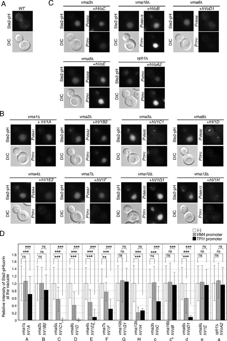
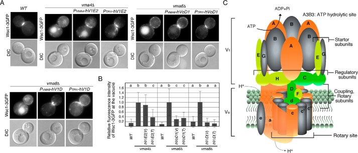
Vacuolar-type H+-ATPase (V-ATPase) is a highly conserved proton pump responsible for acidification of intracellular organelles and potential drug target. It is a multisubunit complex comprising a cytoplasmic V1 domain responsible for ATP hydrolysis and a membrane-embedded Vo domain that contributes to proton translocation across the membrane. Saccharomyces cerevisiae V-ATPase is composed of 14 subunits, deletion of any one of which results in well-defined growth defects. As the structure of V-ATPase and the function of each subunit have been well-characterized in yeast, this organism has been recognized as a preferred model for studies of V-ATPases. In this study, to assess the functional relatedness of the yeast and human V-ATPase subunits, we investigated whether human V-ATPase subunits can complement calcium- or pH-sensitive growth, acidification of the vacuolar lumen, assembly of the V-ATPase complex, and protein sorting in yeast mutants lacking the equivalent yeast genes. These assessments revealed that 9 of the 13 human V-ATPase subunits can partially or fully complement the function of the corresponding yeast subunits. Importantly, sequence similarity was not necessarily correlated with functional complementation. We also found that besides all Vo domain subunits, the V1 F subunit is required for proper assembly of the Vo domain at the endoplasmic reticulum. Furthermore, the human H subunit fully restored the level of vacuolar acidification, but only partially rescued calcium-sensitive growth, suggesting a specific role of the H subunit in V-ATPase activity. These findings provide important insights into functional homologies between yeast and human V-ATPases.

Keywords: Saccharomyces cerevisiae; human; vacuolar ATPase; vacuole; yeast genetics.


Conflict of interest statement

The authors declare that they have no conflicts of interest with the contents of this article

Figures

Figure 1.
Figure 1.
Overview of the experimental design for the complementation test. A, yeast mutants, in which gene encoding each V-ATPase subunit was replaced with the KanMX marker, were obtained from GE Dharmacon. For expression of human genes, two different promoters, the promoter of yeast equivalent genes (PVMA) and the yeast triose-phosphate isomerase gene promoter (PTPI1), were utilized. After expressing the human gene in a mutant lacking the equivalent yeast gene, the following four cell phenotypes, 1) growth ability, 2) vacuolar acidification, 3) localization of the V1 and Vo subunits, and 4) endocytic recycling of Wsc1–3GFP, were evaluated. B, immunoblots showing the expression of GFP-tagged Vma1p and its human equivalent subunit (ATP6V1A) from the VMA1 gene or the TPI1 gene promoter in the vma1Δ mutants. 10 μg of whole-cell extracts from each strain were loaded per lane and immunoblotted with an anti-GFP antibody or anti-glyceraldehyde-3-phosphate dehydrogenase antibody. The bar graphs represent the relative expression levels of these proteins. Data show the mean ± S.D. of three experiments. **, p value < 0.01, one-way ANOVA with Tukey's test. n.s., not statistically significant.
Figure 2.
Figure 2.
Growth complementation assays of yeast V-ATPase (V1 subunit) mutants by the equivalent human gene. A and B, growth assays of yeast V-ATPase (V1 subunit) mutants expressing the human equivalent genes from the native promoter (A) or the TPI1 gene promoter (B). 10-Fold dilutions of cells were spotted on YPD plates containing 0, 30, and 100 mm CaCl2 or pH 7.0. YPD plates were buffered with 50 mm KH2PO4 and 50 mm K2HPO4 and incubated at 25 °C for 72 h. The white dashed line shows boundaries in the composite images. WT and mutant cells in a composite image were spotted and grown on the same plate. After imaging the plate, images of cells spotted in a distant place were cropped separately and combined to create a composite image.
Figure 3.
Figure 3.
Growth complementation assays of yeast V-ATPase (Vo subunit) mutants by the equivalent human gene. A and B, growth assays of yeast V-ATPase (Vo subunit) mutants expressing the human equivalent genes from the native promoter (A) or the TPI1 gene promoter (B). 10-Fold dilutions of cells were spotted on YPD plates containing 0, 30, and 100 mm CaCl2 or 4 mm ZnCl2, pH 7.0, YPD plates were buffered with 50 mm KH2PO4 and 50 mm KH2PO4 and incubated at 25 °C for 72 h.
Figure 4.
Figure 4.
Functional complementation of yeast V-ATPase subunit mutants by the equivalent human gene. A–C, localization and fluorescence of Ste2–pHluorin in WT (A), yeast V-ATPase V1 subunit mutants (B), and Vo subunit mutants (C) expressing the human equivalent genes. Cells expressing Ste2–pHluorin were grown to early logarithmic phase in YPD medium at 25 °C and observed by epifluorescence and differential interference contrast (DIC) microscopy. The pHluorin is sensitive in pH and loses its fluorescence when transported to the acidic vacuole. D, relative intensities of Ste2–pHluorin at the vacuole in yeast V-ATPase mutants. The fluorescence intensity of vacuoles in the vma mutants, including vhp1Δ was calculated by subtracting the average cytosolic background signal from the fluorescence intensity of the individual vacuole. For the relative fluorescence intensity in mutants, the average fluorescence intensity of the vacuole in mutant cells was divided by the average fluorescence intensity of that in WT cells (n = 50). The fluorescence intensities were analyzed by using the program ImageJ Version 1.44. Data show the mean ± standard deviation (S.D.). **, p value < 0.001; ***, p value < 0.0001, one-way ANOVA with Tukey's test. n.s., not statistically significant.
Figure 5.
Figure 5.
Localization of Vo subunit in yeast V-ATPase subunit mutants expressing the equivalent human gene. A and B, localization of Vph1–GFP in yeast V-ATPase V1 subunit mutants (A) and Vo subunit mutants (B) expressing the human equivalent genes. Cells expressing Vph1–GFP and HDEL–mCherry (mCH) were grown to early logarithmic phase in YPD medium at 25 °C and observed by epifluorescence and differential interference contrast (DIC) microscopy. All images were taken with 1-s exposure time under the same conditions. C, quantification of localization of Vph1–GFP. Data show the mean of at least two experiments, with >50 Vph1–GFP-labeled cells counted per experiment (green, vacuolar membrane (Vac); yellow, vacuolar membrane and endoplasmic reticulum (Vac. and ER); red, ER including intravacuolar localization). D, localization of GFP-tagged human VoC protein. Cells expressing GFP-tagged protein and HDEL–mCherry (mCH) were grown and observed as described above. E and F, localization and expression of V5-tagged human VoC protein in the vma3Δ mutant. E, cells were grown to early logarithmic phase, fixed with paraformaldehyde, converted to spheroplasts, and stained with anti-V5 tag antibody. V5-tagged proteins were subsequently visualized with Alexa Fluor 488-conjugated anti-mouse IgG. F, 10 μg of whole-cell extracts from each strain were loaded per lane and immunoblotted with an anti-V5 tag antibody.
Figure 6.
Figure 6.
Localization of V1 subunit in yeast V-ATPase subunit mutants expressing the equivalent human gene. A–C, localization of Vma1–GFP or Vma2–GFP in WT (A), yeast V-ATPase V1 subunit mutants (B), and Vo subunit mutants (C) expressing the human equivalent genes. Cells expressing Vma1–GFP or Vma2–GFP were grown to early logarithmic phase in YPD medium at 25 °C and observed by epifluorescence and differential interference contrast (DIC) microscopy. D, quantification of localization of Vma1–GFP or Vma2–GFP. Data show the mean of at least two experiments, with >50 Vph1–GFP-labeled cells counted per experiment (green, vacuolar membrane (Vac); yellow, vacuolar membrane and cytosol (Vac and Cyt); red, cytosol (Cyt)).
Figure 7.
Figure 7.
Recycling of Wsc1–3GFP in yeast V-ATPase subunit mutants expressing the equivalent human gene. A, localization of Wsc1–3GFP in WT and vma mutants. Cells expressing Wsc1–3GFP were grown to early logarithmic phase in YPD medium at 25 °C and observed by epifluorescence and differential interference contrast (DIC) microscopy. B, quantification of the fluorescence intensity of Wsc1–3GFP at the vacuole in WT and mutant cells. The fluorescence intensity of Wsc1–3GFP at the vacuole was calculated by subtracting average fluorescence intensity in the cytosol from that in the vacuole. For the relative fluorescence intensity in mutants, the average fluorescence intensity in mutant cells was divided by the average fluorescence intensity in WT cells. Data show the mean ± S.D., with >50 cells counted for each strain. Different letters indicate significant difference at p < 0.01 (one-way ANOVA with Tukey's post-hoc test). C, schematic representation of the subunit structure of yeast V-ATPase (2, 28). The subunits whose function were mostly complemented by the corresponding human subunits are shown in green, partially complemented are shown in yellow, and slightly complemented are shown in orange. The subunits that were not complemented are shown in gray.

References

    1. Kane P. M. (2006) The where, when, and how of organelle acidification by the yeast vacuolar H+-ATPase. Microbiol. Mol. Biol. Rev. 70, 177–191 10.1128/MMBR.70.1.177-191.2006 - DOI - PMC - PubMed
    1. Forgac M. (2007) Vacuolar ATPases: rotary proton pumps in physiology and pathophysiology. Nat. Rev. Mol. Cell Biol. 8, 917–929 10.1038/nrm2272 - DOI - PubMed
    1. Mellman I., Fuchs R., and Helenius A. (1986) Acidification of the endocytic and exocytic pathways. Annu. Rev. Biochem. 55, 663–700 10.1146/annurev.bi.55.070186.003311 - DOI - PubMed
    1. Anderson R. G., and Pathak R. K. (1985) Vesicles and cisternae in the trans Golgi apparatus of human fibroblasts are acidic compartments. Cell 40, 635–643 10.1016/0092-8674(85)90212-0 - DOI - PubMed
    1. Mellman I. (1996) Endocytosis and molecular sorting. Annu. Rev. Cell Dev. Biol. 12, 575–625 10.1146/annurev.cellbio.12.1.575 - DOI - PubMed

Publication types

MeSH terms

Substances