. 2019 Jun 14;294(24):9377-9389.
doi: 10.1074/jbc.RA118.006336. Epub 2019 Apr 5.

An unbiased in vitro screen for activating epidermal growth factor receptor mutations

Affiliations

An unbiased in vitro screen for activating epidermal growth factor receptor mutations

Deepankar Chakroborty et al. J Biol Chem. .

Abstract

Cancer tissues harbor thousands of mutations, and a given oncogene may be mutated at hundreds of sites, yet only a few of these mutations have been functionally tested. Here, we describe an unbiased platform for the functional characterization of thousands of variants of a single receptor tyrosine kinase (RTK) gene in a single assay. Our in vitroscreen for activating mutations (iSCREAM) platform enabled rapid analysis of mutations conferring gain-of-function RTK activity promoting clonal growth. The screening strategy included a somatic model of cancer evolution and utilized a library of 7,216 randomly mutated epidermal growth factor receptor (EGFR) single-nucleotide variants that were tested in murine lymphoid Ba/F3 cells. These cells depend on exogenous interleukin-3 (IL-3) for growth, but this dependence can be compensated by ectopic EGFR overexpression, enabling selection for gain-of-function EGFR mutants. Analysis of the enriched mutants revealed EGFR A702V, a novel activating variant that structurally stabilized the EGFR kinase dimer interface and conferred sensitivity to kinase inhibition by afatinib. As proof of concept for our approach, we recapitulated clinical observations and identified the EGFR L858R as the major enriched EGFR variant. Altogether, iSCREAM enabled robust enrichment of 21 variants from a total of 7,216 EGFR mutations. These findings indicate the power of this screening platform for unbiased identification of activating RTK variants that are enriched under selection pressure in a model of cancer heterogeneity and evolution.

Keywords: ERBB; cancer; cancer biology; clonal selection; directed evolution; driver mutations; epidermal growth factor receptor (EGFR); mutagenesis in vitro; oncogene; passenger mutations; somatic evolution; targeted therapies; tyrosine kinase inhibitors; tyrosine-protein kinase (tyrosine kinase).


Conflict of interest statement

P. A. Jänne has received consulting fees from AstraZeneca, Boehringer Ingelheim, Pfizer, Merrimack Pharmaceuticals, Roche/Genentech, Chugai Pharmaceuticals, ACEA Biosciences, and Ariad Pharmaceuticals and sponsored research funding from Astellas Pharmaceuticals, AstraZeneca, Daiichi Sankyo, and PUMA and receives post-marketing royalties on DFCI-owned intellectual property on EGFR mutations licensed to Lab Corp. K. Elenius has a research agreement with Boehringer Ingelheim and ownership interest in Abomics, Orion, and Roche

Figures

Figure 1.
Figure 1.
Cancer-associated EGFR mutations. The y axis shows the number of EGFR mutations reported in different cancer types in the COSMIC database (version 86) (http://cancer.sanger.ac.uk/cosmic).8 Please note that the y axis is shown in log10 scale. The total number of EGFR mutations in the database was 21,339. The x axis shows the position of the mutated residue in the EGFR primary sequence. Mutation hot spots, such as L858R and exon 19 deletions, are indicated with labels.
Figure 2.
Figure 2.
iSCREAM pipeline for identification of driver mutations. An expression library including random EGFR mutations was generated by amplifying WT human EGFR cDNA using error-prone PCR and cloning the amplicons into a retroviral mammalian expression vector. The library was subsequently transduced into IL-3–dependent Ba/F3 cells that can survive IL-3 withdrawal if expressing an activated EGFR variant. Upon IL-3 deprivation, clones of Ba/F3 cells expressing WT EGFR or passenger mutations die, whereas clones expressing activating EGFR mutants survive and proliferate. By analyzing the EGFR insert in the surviving cell population as well as in the original library used for infection using targeted next-generation sequencing, it can be determined which mutations are enriched along with the clonal expansion and thus functionally driving the growth.
Figure 3.
Figure 3.
Clonal selection of activating EGFR mutations. The scatter blot demonstrates the enrichment of few specific EGFR mutations in Ba/F3 cells engineered to express 7,216 EGFR variants and to grow in an EGFR activity–dependent manner. The y axis indicates the -fold change of the variant frequency after growing the cells for 2 weeks when compared with the variant frequency in the original mutation library used to transduce the cells. The x axis indicates the position of the mutated residue on the EGFR primary sequence. The size of the dot as well as the intensity of its red color indicate the relative variant frequency of the mutation in the growing cell pool at the 2-week time point. The dashed horizontal line indicates the -fold change level (24.5) for statistically significant difference from the normal distribution after log2 transformation (q < 0.0001).
Figure 4.
Figure 4.
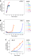
EGFR mutations L858R, T790M, and A702V promote ligand-independent growth. Ba/F3 cells were infected with lentiviral vectors encoding the indicated EGFR variants and cultured in the presence (A) or absence (B and C) of exogenous IL-3. Cell viability was measured by MTT analyses. A, all cell lines were viable in the presence of IL-3. B, after initial withdrawal of IL-3, cells expressing activating mutations emerged as IL-3–independent after a refractory phase of 7–14 days. C, after initial establishment of IL-3 independence, cells harboring activating mutants readily proliferated in the absence of IL-3. Error bars, S.D.
Figure 5.
Figure 5.
Induction of EGFR expression upon EGFR addiction. A, a Western blot analysis of the indicated cell lines cultured in the presence or absence of IL-3. Two independent clones of cells expressing the A702V variant were analyzed (lanes 4 and 5). B, Western analyses of the indicated Ba/F3 cell lines maintained in the presence of IL-3 and stimulated or not with 10 ng/ml EGF for 10 min. C and D, flow cytometry analyses of the expression of EGFR in the indicated cell lines. The three activating EGFR mutants were analyzed both after maintaining them in the presence (C) or absence (D) of IL-3. E, real-time RT-PCR analysis of EGFR expression in the Ba/F3 cell lines expressing the indicated EGFR variants after maintaining them in the presence or absence of IL-3. Three data points and their means (horizontal lines) are indicated.
Figure 6.
Figure 6.
Effect of EGFR A702V on kinase interface. Modeled A702V (receiver kinase) interaction with Ile-941 (activator kinase) at the asymmetric dimer interface in the 2.6 Å resolution EGFR X-ray structure with bound ATP peptide analogue (PDB code 2GS6). A structural water (small red sphere) is nearby. Carbon atoms for the Ile-941 and A702V are shown as orange and magenta spheres, respectively; the red and blue spheres, where seen, correspond to main-chain carbonyl oxygen and amino groups, respectively.
Figure 7.
Figure 7.
Variable drug sensitivity of activating EGFR mutations. A, drug response curves of Ba/F3 cells expressing the indicated EGFR variants or EGFP (vector control). Cells were cultured for 3 days in the presence of the indicated concentrations of cetuximab, erlotinib, or afatinib. Cells expressing EGFR variants were cultured in the absence of IL-3, and vector control cells were cultured in the presence of IL-3. B, Western blotting analyses of EGFR phosphorylation at Tyr-1110 in Ba/F3 cells expressing the indicated EGFR variants after treating the cells for 3 h with the indicated concentrations of erlotinib or afatinib. Error bars, S.D.

References

    1. Lawrence M. S., Stojanov P., Polak P., Kryukov G. V., Cibulskis K., Sivachenko A., Carter S. L., Stewart C., Mermel C. H., Roberts S. A., Kiezun A., Hammerman P. S., McKenna A., Drier Y., Zou L., et al. (2013) Mutational heterogeneity in cancer and the search for new cancer-associated genes. Nature 499, 214–218 10.1038/nature12213 - DOI - PMC - PubMed
    1. Zehir A., Benayed R., Shah R. H., Syed A., Middha S., Kim H. R., Srinivasan P., Gao J., Chakravarty D., Devlin S. M., Hellmann M. D., Barron D. A., Schram A. M., Hameed M., Dogan S., et al. (2017) Mutational landscape of metastatic cancer revealed from prospective clinical sequencing of 10,000 patients. Nat. Med. 23, 703–713 10.1038/nm.4333 - DOI - PMC - PubMed
    1. Hyman D. M., Taylor B. S., and Baselga J. (2017) Implementing genome-driven oncology. Cell 168, 584–599 10.1016/j.cell.2016.12.015 - DOI - PMC - PubMed
    1. Vogelstein B., Papadopoulos N., Velculescu V. E., Zhou S., Diaz L. A. Jr., and Kinzler K. W. (2013) Cancer genome landscapes. Science 339, 1546–1558 10.1126/science.1235122 - DOI - PMC - PubMed
    1. Chang M. T., Asthana S., Gao S. P., Lee B. H., Chapman J. S., Kandoth C., Gao J., Socci N. D., Solit D. B., Olshen A. B., Schultz N., and Taylor B. S. (2016) Identifying recurrent mutations in cancer reveals widespread lineage diversity and mutational specificity. Nat. Biotechnol. 34, 155–163 10.1038/nbt.3391 - DOI - PMC - PubMed

Publication types