. 2019 Jun 3;218(6):1787-1798.
doi: 10.1083/jcb.201811139. Epub 2019 Apr 5.

ATG2 transports lipids to promote autophagosome biogenesis

Affiliations

ATG2 transports lipids to promote autophagosome biogenesis

Diana P Valverde et al. J Cell Biol. .

Abstract

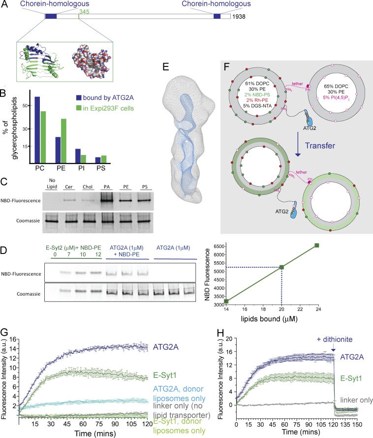
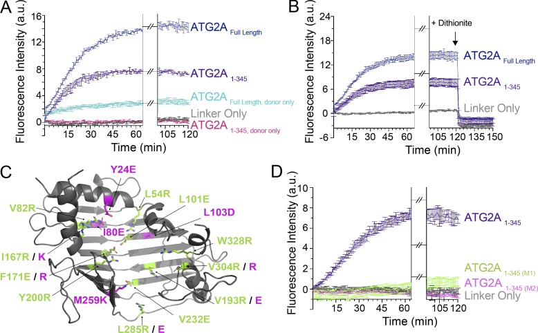
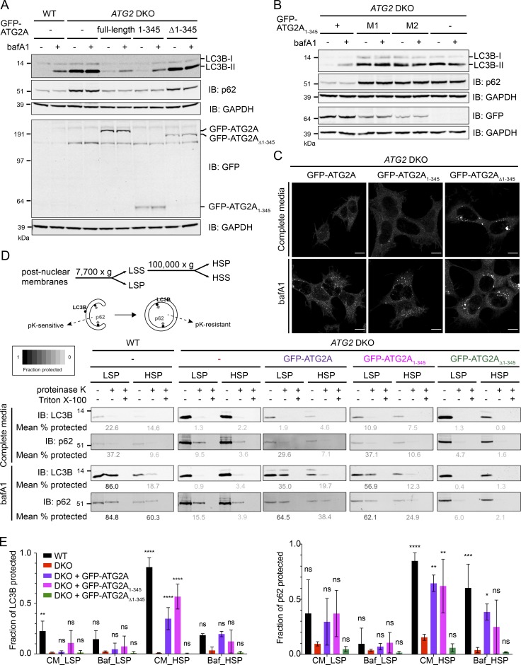
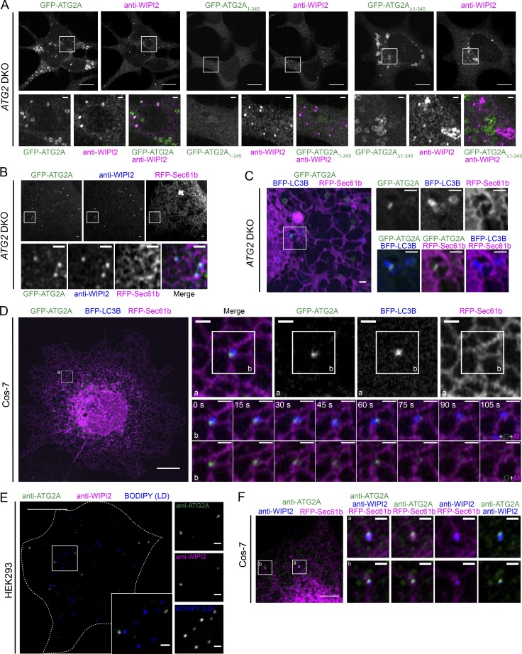
During macroautophagic stress, autophagosomes can be produced continuously and in high numbers. Many different organelles have been reported as potential donor membranes for this sustained autophagosome growth, but specific machinery to support the delivery of lipid to the growing autophagosome membrane has remained unknown. Here we show that the autophagy protein, ATG2, without a clear function since its discovery over 20 yr ago, is in fact a lipid-transfer protein likely operating at the ER-autophagosome interface. ATG2A can bind tens of glycerophospholipids at once and transfers lipids robustly in vitro. An N-terminal fragment of ATG2A that supports lipid transfer in vitro is both necessary and fully sufficient to rescue blocked autophagosome biogenesis in ATG2A/ATG2B KO cells, implying that regulation of lipid homeostasis is the major autophagy-dependent activity of this protein and, by extension, that protein-mediated lipid transfer across contact sites is a principal contributor to autophagosome formation.


Figures

Figure 1.
Figure 1.
ATG2A binds and transfers glycerophospholipids between membranes. (A) ATG2 architecture. Sequences homologous to VPS13 proteins are indicated. Inset shows a fragment from the ATG2A N terminus modeled on the crystal structure of Vps13 (PDBID 6CBC). The chorein_N sequence is indicated in blue. A space-filling model colored according to atom type (red for oxygen, blue for nitrogen, and white for carbon) suggests that a cavity in this fragment is hydrophobic and suitable for solubilizing glycerophospholipid fatty acid chains. (B) Lipids that copurified with ATG2A from Expi293 cells according to abundance. No sterols, diglycerides, or triglycerides were detected. Relative abundance of glycerophospholipids in Expi293 cells is indicated (Lees et al., 2017). (C) ATG2A was incubated with NBD-tagged lipids and examined by native PAGE. Phospholipids, visualized by their fluorescence, comigrated with protein, visualized by Coomassie blue staining. Cer, ceramide; Chol, cholesterol; PA, phosphatidic acid. (D) A native gel assay was used to compare NBD fluorescence associated with ATG2A and indicated quantities of the extended-synaptotagmin2 (E-Syt2) SMP domain, known to accommodate two glycerophospholipids within its cavity (Schauder et al., 2014). Based on this comparison, each ATG2A binds to ∼20 lipid molecules. The experiment was performed in triplicate. SD is shown. (E) The 3D cryo-EM reconstruction of ATG2A at a nominal resolution of ∼15 Å, shown in mesh representation (6.5 signal/noise). A cavity (or cavities) highlighted blue runs along the length of ATG2A. Fig. S1 shows additional views of ATG2A. (F) In the transfer assay, donor and acceptor liposomes (compositions indicated) were tethered together in the presence or absence ATG2A linked to the donor liposomes. The assay monitors the increase in NBD-PS fluorescence after lipid transfer from donor liposomes, where NBD fluorescence is quenched via FRET with Rh-PE, to acceptor liposomes. (G) The fluorescence increase observed is consistent with lipid transfer at a rate similar to the lipid transfer domain of extended-synaptotagmin1 (E-Syt1), a previously validated lipid transporter (Saheki et al., 2016; Yu et al., 2016; Bian et al., 2018), tethered analogously to ATG2A. The fluorescence increase is much smaller when only donor but not acceptor liposomes are present. The small but still significant increase under these conditions is most likely due to lipid extraction by the transport proteins and would not be expected in the case of fusion or hemifusion between donor liposomes. The increase in this case is larger for ATG2A than E-Syt1, likely reflecting that the ATG2A cavity accommodates more lipids. All experiments were performed in triplicate. SD is shown. (H) Addition of dithionite reduces the NBD fluorescence equally for ATG2A, E-Syt1, or the tether-only control, indicating that membrane fusion has not occurred. Each experiment was performed in triplicate; SDs are shown.
Figure 2.
Figure 2.
The N-terminal module in ATG2A transfers lipids in vitro. (A) FRET-based lipid transfer assay (as in Fig. 1 G) comparing transfer or lipid extraction only (no acceptor liposomes) by full-length and mini-ATG2A. The higher plateaus for full-length ATG2A likely reflect a more extensive cavity accommodating more lipids. (B) The dithionite assay (as in Fig. 1 H) rules out that the increases in NBD fluorescence are due to liposome fusion. (C) In two mutant forms of mini-ATG2 (M1 and M2), the hydrophobic cleft was made hydrophilic by the inclusion of charged amino acids, indicated in green and purple, respectively, on the structural model. (D) Both mutants lose the capacity to transfer lipids in the FRET-based assay. Each experiment was performed in triplicate; SDs are shown.
Figure 3.
Figure 3.
The N-terminal module in ATG2A is both required and sufficient for autophagosome maturation in ATG2 DKO cells. (A) Immunoblots (IB) of WT, ATG2 DKO, and DKO cells stably expressing full-length GFP-ATG2A, GFP-ATG2A1–345, or GFP-ATG2AΔ1–345, grown in complete medium ± bafA1. Normal autophagy flux is indicated by the increase in LC3B-II when lysosomes are poisoned with bafA1 (WT, full-length rescue and 1–345 rescue). Likewise, p62 levels are low in autophagy-competent cells. Flux is blocked in the DKO cells or cells rescued with Δ1–345. Quantification of autophagy rescues are shown in Fig. S2 F. GAPDH is a loading control. (B) Immunoblots of ATG2 DKO cells rescued with the minifragment or either of the two mutant clusters described in Fig. 2. Quantification of p62 levels is shown in Fig. S2 J. (C) Anti-LC3B IF images of DKO cells stably expressing GFP-ATG2A constructs in A and grown in complete medium or complete medium with bafA1. Cells lacking a functional ATG2A (GFP-ATG2AΔ1–345), accumulate large LC3-positive structures, while expression of functional ATG2 (GFP-ATG2A and GFP-ATG2A1–345) leads to WT-like small puncta that accumulate under bafA1 treatment. Scale bars: 10 µm. (D) Postnuclear membranes from indicated cell lines grown in complete medium were differentially centrifuged to generate LSP and HSP fractions. Each fraction was treated with proteinase K, with and without Triton X-100, and subjected to anti-LC3B and anti-p62 immunoblotting. Percentage of protected LC3B or p62 was calculated from the ratio of signal in the proteinase K–treated lane over the nontreated control. Cells with functional ATG2A accumulate proteinase-K resistant LC3B and p62, particularly in the LSP (WT, GFP-ATG2A, GFP-ATG2A1–345), while autophagy-impaired cells (DKO and GFP-ATG2AΔ1–345) do not. The mean value of three independent experiments is shown below. (E) Full quantification of p62 and LC3-II levels from experiments in D. Statistical significance was calculated by two-way ANOVA. *, P < 0.05; **, P < 0.01; ***, P < 0.001; ****, P < 0.0001. Expression levels of all GFP-ATG2A constructs is shown in Fig. S2 I. ns, not significant.
Figure 4.
Figure 4.
Overexpressed GFP-ATG2A and endogenous ATG2A localizes at ER–autophagosomal membrane contact sites. (A) ATG2 DKO HEK293 cells stably expressing the indicated GFP-ATG2A construct were treated with OA to enlarge LDs, and then incubated in starvation medium (Earle’s balanced salt solution) and subjected to anti-WIPI2 IF. Greater than 88% of all WIPI2 positive structures are also GFP-ATG2A positive. Targeting to autophagosomes (WIPI2 structures) is lost with removal of the mini-ATG2 sequence (GFP-ATG2AΔ1–345). Scale bars: 10 µm; zoomed field: 1 µm. (B) GFP-ATG2A localized with early autophagosome markers is also associated with the ER. Anti-WIPI2 IF confocal images in starved ATG2 DKO cells stably expressing GFP-ATG2A and transiently expressing RFP-Sec61b. Scale bars: 1 µm. (C) The late autophagosome marker LC3B colocalized with GFP-ATG2A ∼13% of the time, and these LC3B/GFP-ATG2-positive puncta consistently also associated with the ER. Live imaging of starved ATG2 DKO cells stably expressing GFP-ATG2A, transiently expressing RFP-Sec61b and BFP-LC3B, and incubated with OA. Note that ring-like GFP-ATG2 structures are likely LDs, are not LC3B-positive, and are not restricted to the ER. Full time course in Fig. S3 B indicates GFP-ATG2A remains directly associated with the ER during the entire time it localizes to the autophagosome. Scale bars: 1 µm. (D) Time-lapse imaging of starved Cos-7 cells stably expressing GFP-ATG2A and transiently expressing RFP-Sec61b and BFP-LC3B. ER localization of autophagosome-associated GFP-ATG2A is similar in WT Cos-7 cells as in DKO HEK293 cells, and again persists throughout the residence time of GFP-ATG2A at LC3B-positive structures. A second longer video example is shown in Fig. S3 C. Scale bars: 10 µm; zoomed field: 1 µm. (E) Endogenous ATG2A is localized only on autophagosomes (WIPI2-positive) and not LDs. Commercially available ATG2A antibodies were precleared with ATG2 DKO cellular material to improve signal to noise (Fig. S3). Then, starved HEK293 cells were treated with OA, labeled with BODIPY 558/568, and subjected to anti-WIPI2 and precleared anti-ATG2A IF. Scale bars: 10 µm; zoomed field: 1 µm. (F) Endogenous ATG2A is associated with autophagosome markers and the ER. Starved Cos-7 cells were transiently transfected with RFP-Sec61b, treated with OA, and subjected to anti-WIPI2 and precleared anti-ATG2A IF. Scale bars: 5 µm; zoomed field: 1 µm. For all panels, representative confocal images from at least three independent experiments are shown.

Comment in

References

    1. Axe E.L., Walker S.A., Manifava M., Chandra P., Roderick H.L., Habermann A., Griffiths G., and Ktistakis N.T.. 2008. Autophagosome formation from membrane compartments enriched in phosphatidylinositol 3-phosphate and dynamically connected to the endoplasmic reticulum. J. Cell Biol. 182:685–701. 10.1083/jcb.200803137 - DOI - PMC - PubMed
    1. Bian X., Saheki Y., and De Camilli P.. 2018. Ca2+ releases E-Syt1 autoinhibition to couple ER-plasma membrane tethering with lipid transport. EMBO J. 37:219–234. 10.15252/embj.201797359 - DOI - PMC - PubMed
    1. Chowdhury S., Otomo C., Leitner A., Ohashi K., Aebersold R., Lander G.C., and Otomo T.. 2018. Insights into autophagosome biogenesis from structural and biochemical analyses of the ATG2A-WIPI4 complex. Proc. Natl. Acad. Sci. USA. 115:9792–9801. 10.1073/pnas.1811874115 - DOI - PMC - PubMed
    1. Fang X.T., Sehlin D., Lannfelt L., Syvänen S., and Hultqvist G.. 2017. Efficient and inexpensive transient expression of multispecific multivalent antibodies in Expi293 cells. Biol. Proced. Online. 19:11 10.1186/s12575-017-0060-7 - DOI - PMC - PubMed
    1. Gómez-Sánchez R., Rose J., Guimarães R., Mari M., Papinski D., Rieter E., Geerts W.J., Hardenberg R., Kraft C., Ungermann C., and Reggiori F.. 2018. Atg9 establishes Atg2-dependent contact sites between the endoplasmic reticulum and phagophores. J. Cell Biol. 217:2743–2763. 10.1083/jcb.201710116 - DOI - PMC - PubMed

Publication types

MeSH terms

Associated data