Review
. 2019 Apr 5;4(34):eaas9673.
doi: 10.1126/sciimmunol.aas9673.

Location, location, location: Tissue resident memory T cells in mice and humans

Affiliations
Review

Location, location, location: Tissue resident memory T cells in mice and humans

Peter A Szabo et al. Sci Immunol. .

Abstract

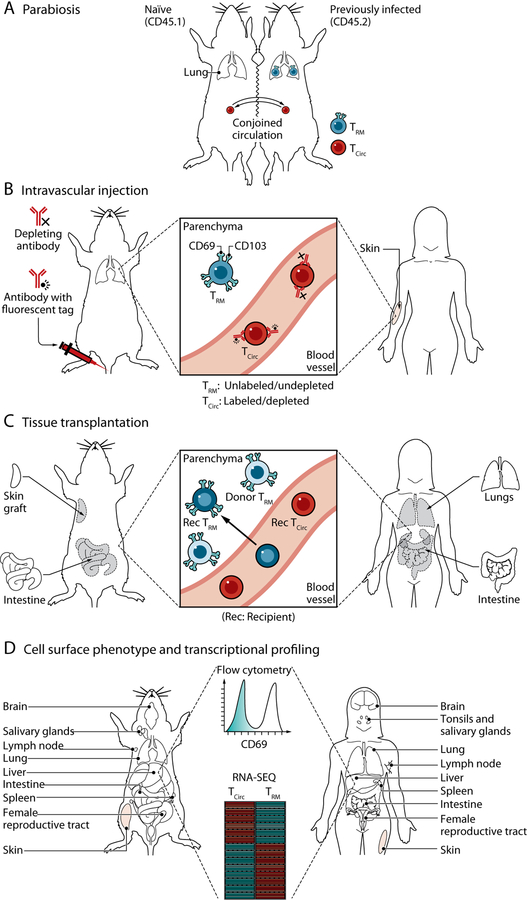
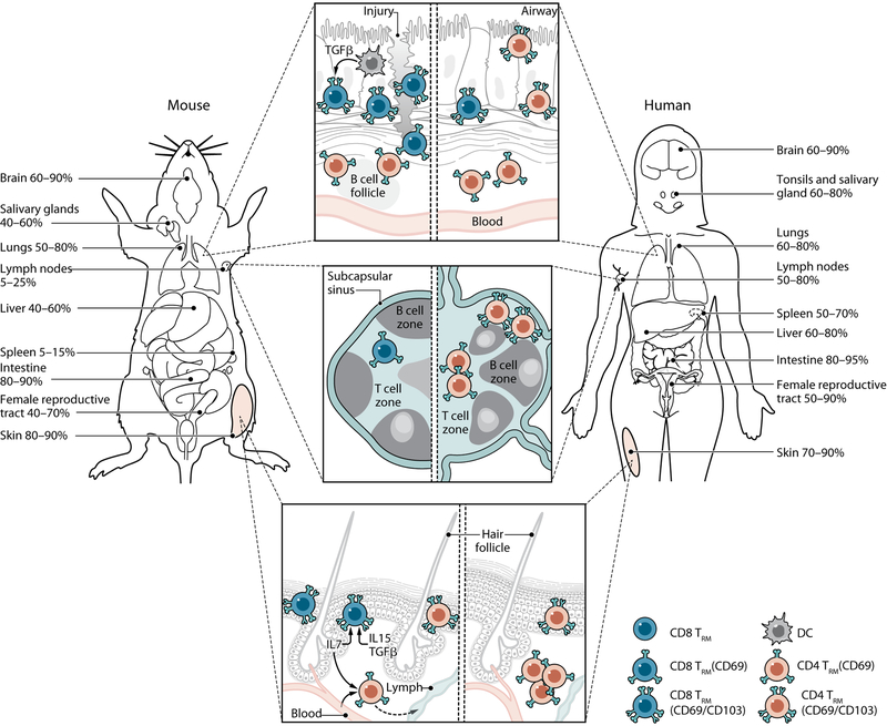
The discovery of T cells resident in diverse tissues has altered our understanding of adaptive immunity to encompass site-specific responses mediated by tissue-adapted memory T cells throughout the body. Here, we discuss the key phenotypic, transcriptional, and functional features of these tissue-resident memory T cells (TRM) as established in mouse models of infection and translated to humans by novel tissue sampling approaches. Integration of findings from mouse and human studies may hold the key to unlocking the potential of TRM for promoting tissue immunity and preventing infection.


Figures

Fig. 1.
Fig. 1.. Approaches for the study of TRM in mice and humans.
(A) Parabiosis. Parabiotic surgery, shown here as the conjoining of the skin between two congenic mice, results in anastomosis of the vasculature assess in vivo circulatory potential. TRM (generated by previous infection in one of the mice) fail to equilibrate between parabionts, providing evidence for tissue residency. (B) In vivo antibody labeling. Intravascular injection of fluorescently labeled or depleting antibodies targets cells in vascular compartments, leaving those outside of the circulation (within the tissue parenchyma) protected from antibody labeling. (C) Tissue transplantation. Analysis of donor and recipient T cells within transplanted organs and tissues allow assessment of retention of donor T cells within (and outside) the graft and influx of recipient cells from circulation. (D) Cell phenotype and transcriptional profile. Expression of CD69, CD103, and other TRM accessory markers (Table 1) by means of flow cytometry is frequently used to define TRM in human tissues or mouse models of infection. Transcriptional profiling (such as microarray or RNA-sequencing) has been used to define gene signatures and TFs associated with tissue residency. Credit: A Kitterman/Science Immunology
Fig. 2.
Fig. 2.. TRM compartmentalization and tissue niches in mice and humans.
Noncirculating TRM take up residence in distinct mucosal, lymphoid, and barrier sites throughout mice and human tissues. Percentages in each tissue denote the frequency of T cells that exhibit surface markers indicating residency (CD69 +/−CD103) in both humans and mouse models of infection with references indicated. (Top) In mice, CD103+CD8+ TRM accumulate in within the lung epithelium or areas of tissue injury and are maintained by DCs producing TGF-β. CD4+TRM localize around B cell follicles. In humans, CD103+CD8+ TRM accumulate in the epithelium, and CD103CD4+TRM are common in the lamina propria. (Middle) LN TRM are infrequent in conventional specific pathogen–free mice, lack CD103 and reside primarily near the subcapsular sinus. LN CD4+ and CD8+ TRM in humans are more abundant as compared with mice. (Bottom) CD103+CD8+ TRM in previously infected mice are the most prevalent skin TRM population, reside in the epidermis, and are maintained by IL-7, IL-5, and TGF-β signals. A proportion of CD4+ T cells expressing CD69 in the dermis retain their ability to recirculate. In humans, dermal CD103CD4+ TRM are the most prevalent population, whereas CD103+CD8+ TRM predominate in the epidermis. Credit: A Kitterman/Science Immunology

References

    1. Sallusto F, Lenig D, Forster R, Lipp M, Lanzavecchia A, Two subsets of memory T lymphocytes with distinct homing potentials and effector functions. Nature 401, 708–712 (1999). - PubMed
    1. Masopust D, Vezys V, Marzo AL, Lefrancois L, Preferential localization of effector memory cells in nonlymphoid tissue. Science 291, 2413–2417. (2001). - PubMed
    1. Reinhardt RL, Khoruts A, Merica R, Zell T, Jenkins MK, Visualizing the generation of memory CD4 T cells in the whole body. Nature 410, 101–105. (2001). - PubMed
    1. Wakim LM, Waithman J, van Rooijen N, Heath WR, Carbone FR, Dendritic cell-induced memory T cell activation in nonlymphoid tissues. Science 319, 198–202 (2008). - PubMed
    1. Gebhardt T et al., Memory T cells in nonlymphoid tissue that provide enhanced local immunity during infection with herpes simplex virus. Nat Immunol 10, 524–530 (2009). - PubMed