. 2019 Apr 5;10(1):1580.
doi: 10.1038/s41467-019-09389-2.

Perfluorocarbon regulates the intratumoural environment to enhance hypoxia-based agent efficacy

Affiliations

Perfluorocarbon regulates the intratumoural environment to enhance hypoxia-based agent efficacy

Wenguang Wang et al. Nat Commun. .

Abstract

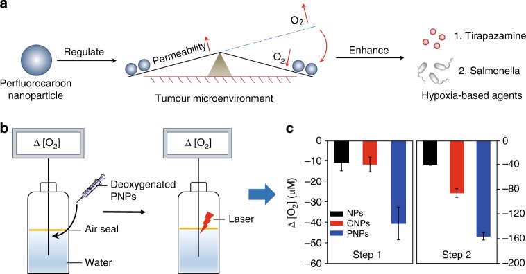
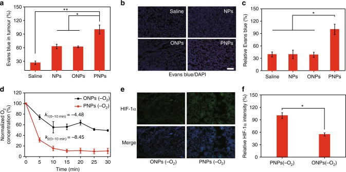
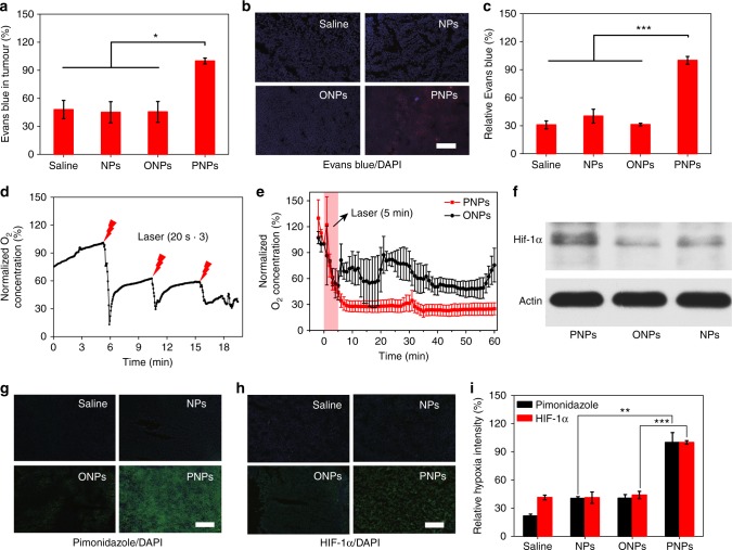
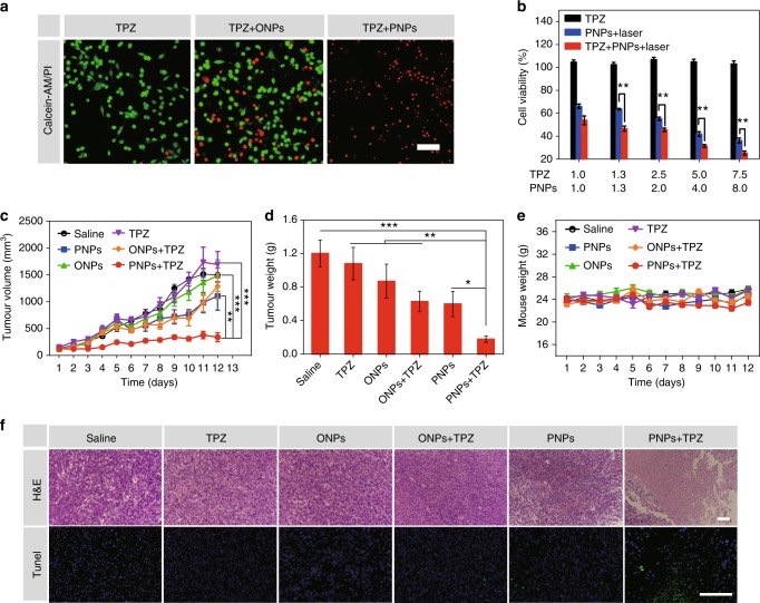
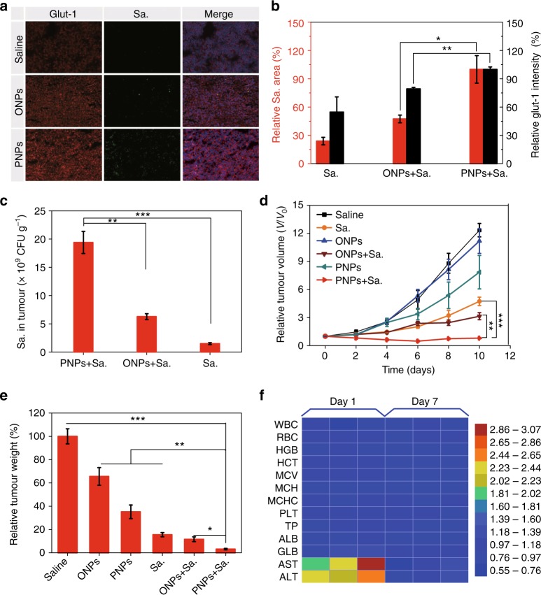
Hypoxia-based agents (HBAs), such as anaerobic bacteria and bioreductive prodrugs, require both a permeable and hypoxic intratumoural environment to be fully effective. To solve this problem, herein, we report that perfluorocarbon nanoparticles (PNPs) can be used to create a long-lasting, penetrable and hypoxic tumour microenvironment for ensuring both the delivery and activation of subsequently administered HBAs. In addition to the increased permeability and enhanced hypoxia caused by the PNPs, the PNPs can be retained to further achieve the long-term inhibition of intratumoural O2 reperfusion while enhancing HBA accumulation for over 24 h. Therefore, perfluorocarbon materials may have great potential for reigniting clinical research on hypoxia-based drugs.


Conflict of interest statement

The authors declare no competing interests.

Figures

Fig. 1
Fig. 1
A schematic diagram of PNP-mediated improvements in the function of HBAs and O2 absorption and consumption by PNPs. a Schematic diagram of PNP-enhanced hypoxia-based drug efficacy. PNPs enable both enhanced tumour permeability and long-term hypoxia to improve the efficacy of HBAs. b Schematic illustration of the measurement of the O2 absorption/consumption ability of the PNPs. Step 1: Deoxygenated samples were added to H2O sealed with oil; changes in DO (dissolved O2, Δ [O2]) were measured using an inserted O2 probe. Step 2: Changes in DO before and after laser irradiation. c Quantitative determination of DO variations in steps 1 and 2 (n = 3). Data are presented as the mean ± s.e.m. Source data are provided as a Source Data file
Fig. 2
Fig. 2
Long-term effects of enhanced permeability and hypoxia mediated by deoxygenated PNPs in vivo. a Analysis of Evans blue accumulation in tumours 24 h after the intravenous injection of deoxygenated PNPs. Evans blue in the whole tumour was extracted and quantified by ultraviolet–visible (UV–vis) spectroscopy (n = 3). b Ex vivo fluorescence images of Evans blue (red) and 4′,6-diamidino-2-phenylindole (DAPI) (blue) staining (scale bar, 100 µm). c Quantification of Evans blue accumulation in b (n = 3). d Changes in DO after the intratumoural injection of deoxygenated PNPs and oil nanoparticles (ONPs). e Representative immunofluorescence images of hypoxia-inducible factor-1α (HIF-1α) (green) staining at 24 h after the intratumoural injection of deoxygenated PNPs and ONPs (scale bar, 100 µm). f Quantification of HIF-1α expression shown in e (n = 4). Data are presented as the mean ± s.e.m. *p< 0.05, **p < 0.01 (unpaired, two-way t tests). Source data are provided as a Source Data file
Fig. 3
Fig. 3
Increased tumour permeability and hypoxia mediated by deoxygenated PNPs under laser irradiation. a The accumulation of Evans blue in tumours was quantified 24 h after the intravenous injection of PNPs and 3 h after laser irradiation (5 min, 808 nm, 400 mW cm−2), (n = 3). b Ex vivo fluorescence images of Evans blue (red) in tumour sections from a. Nuclei were stained with 4′,6-diamidino-2-phenylindole (DAPI) (blue), (scale bar, 100 µm), (n = 3). c Relative quantification of Evans blue accumulation in b (n = 4). d Changes in dissolved oxygen (DO) during three cycles of laser irradiation (808 nm, 2 W cm−2, 20 s) after intratumoural administration. e Changes in DO after laser irradiation (808 nm, 400 mW cm−2) for 5 min at 24 h post-intravenous injection (n = 3). f Western blot of intratumoural hypoxia-inducible factor-1α (HIF-1α) expression at 24 h after laser irradiation. g, h Tumour sections were stained with anti-pimonidazole antibody (green), anti-HIF-1α antibody (green) and DAPI (blue) at 24 h post irradiation (scale bars, 200 µm). i Quantification of pimonidazole and HIF-1α staining (n ≥ 3). Data are presented as the mean ± s.e.m. *p< 0.05, **p < 0.01 and ***p < 0.001 (unpaired, two-way t tests). Source data are provided as a Source Data file
Fig. 4
Fig. 4
PNPs enhanced TPZ chemotherapy. a Fluorescence images of CT26 cells cultured with TPZ and oil nanoparticles (ONPs) or PNPs under laser irradiation (808 nm, 400 mW cm−2). Viable cells were stained with Calcein-AM (green), and dead/late apoptotic cells were stained with propidium iodide (PI) (red), (scale bar, 100μm). b Viability of treated cells (n = 3). c Tumour growth curves of treated mice (n ≥ 6 per group). Both PNPs and TPZ were injected intravenously into mice on days 1, 3 and 5. Tumours in groups 3, 4, 5 and 6 were irradiated three times (808 nm, 400 mW cm−2) for 5 min at 24 h after the administration of the PNPs. d Normalized averages of the tumour weight on day 12. e Changes in body weight. f Haematoxylin and eosin (H&E) (scale bars, 100 µm) and terminal deoxynucleotidyl transferase dUTP nick-end labelling (TUNEL) (green) (scale bars, 100 µm) staining of CT26 tumour sections. Samples were collected from different groups on day 8 post administration. Data are presented as the mean ± s.e.m. *p < 0.05, **p < 0.01, *** p < 0.001 (unpaired, two-way t tests). Source data are provided as a Source Data file
Fig. 5
Fig. 5
PNPs enabled hypoxia-based bacterial cancer therapy. a The hypoxia intensity, Salmonella VNP20009 (Sa.) accumulation and nuclei were determined by staining with anti-glut-1 antibody (red), anti-Sa. antibody (green) and 4′,6-diamidino-2-phenylindole (DAPI) (blue), respectively (scale bar, 100 µm). Sa. (5 × 106 CFU) was intravenously administered to mice 1 h before irradiation. The mice were exposed to the laser (808 nm, 400 mW cm−2) for 5 min at 24 h after they were intravenously injected with PNPs, oil nanoparticles (ONPs) or saline. Immunofluorescence images of tumour sections were analysed on day 3 post irradiation (n ≥ 3). b Semiquantitative analysis of the relative hypoxia intensity (the black y-axis) and Sa. distribution (the red y-axis) in a. c The distribution of Sa. in tumours was also measured by colony-forming assay on day 3 (n = 3). d Tumour growth curves of mice subjected to different treatments (n = 4–9). e Normalized tumour weight at day 10 post treatment. f Haematologic indexes and blood biochemistry of mice that were intravenously injected with VNP20009. The experiment was carried out on days 1 and 7 after the injection of VNP20009 (5 × 106 CFU mouse−1, n = 3) and saline. Values are the mean ± s.e.m. *p< 0.05, **p < 0.01, ***p < 0.001 (unpaired, two-way t tests). Source data are provided as a Source Data file

References

    1. Wilson WR, Hay MP. Targeting hypoxia in cancer therapy. Nat. Rev. Cancer. 2011;11:393–410. doi: 10.1038/nrc3064. - DOI - PubMed
    1. Brown JM, Wilson WR. Exploiting tumour hypoxia in cancer treatment. Nat. Rev. Cancer. 2004;4:437. doi: 10.1038/nrc1367. - DOI - PubMed
    1. Workman P, Stratford IJ. The experimental development of bioreductive drugs and their role in cancer therapy. Cancer Metastas. Rev. 1993;12:73–82. doi: 10.1007/BF00689802. - DOI - PubMed
    1. Brown JM. SR 4233 (tirapazamine): a new anticancer drug exploiting hypoxia in solid tumours. Br. J. Cancer. 1993;67:1163–1170. doi: 10.1038/bjc.1993.220. - DOI - PMC - PubMed
    1. Ganai S, Arenas RB, Sauer JP, Bentley B, Forbes NS. In tumors Salmonella migrate away from vasculature toward the transition zone and induce apoptosis. Cancer Gene. Ther. 2011;18:457. doi: 10.1038/cgt.2011.10. - DOI - PMC - PubMed

Publication types